. 2006 Jul;17(7):3187-96.
doi: 10.1091/mbc.e05-12-1122. Epub 2006 May 10.

The docking protein Cas links tyrosine phosphorylation signaling to elongation of cerebellar granule cell axons

Affiliations

The docking protein Cas links tyrosine phosphorylation signaling to elongation of cerebellar granule cell axons

Jinhong Huang et al. Mol Biol Cell. 2006 Jul.

Abstract

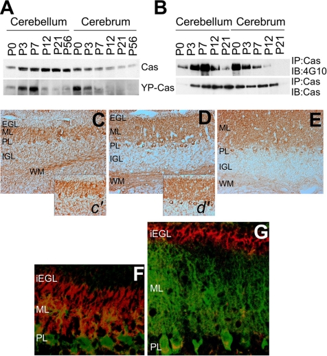
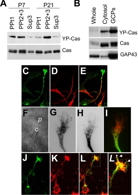
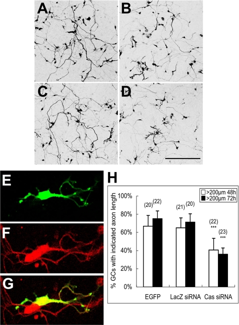
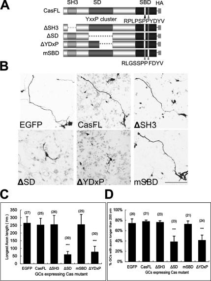
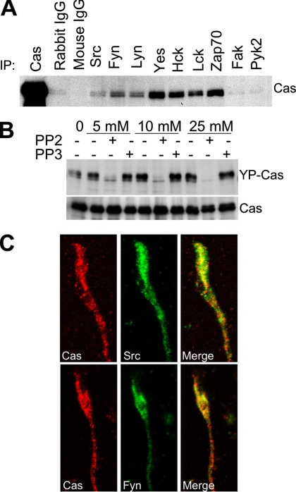
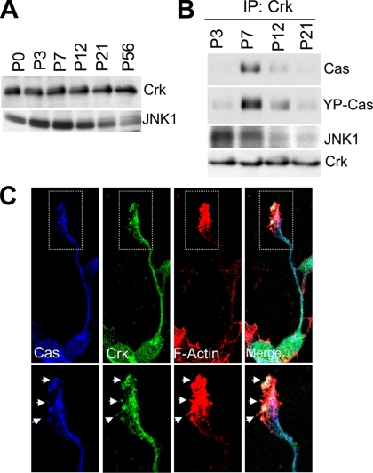
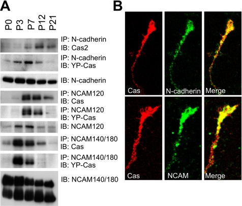
Crk-associated substrate (Cas) is a tyrosine-phosphorylated docking protein that is indispensable for the regulation of the actin cytoskeletal organization and cell migration in fibroblasts. The function of Cas in neurons, however, is poorly understood. Here we report that Cas is dominantly enriched in the brain, especially the cerebellum, of postnatal mice. During cerebellar development, Cas is highly tyrosine phosphorylated and is concentrated in the neurites and growth cones of granule cells. Cas coimmunoprecipitates with Src family protein tyrosine kinases, Crk, and cell adhesion molecules and colocalizes with these proteins in granule cells. The axon extension of granule cells is inhibited by either RNA interference knockdown of Cas or overexpression of the Cas mutant lacking the YDxP motifs, which are tyrosine phosphorylated and thereby interact with Crk. These findings demonstrate that Cas acts as a key scaffold that links the proteins associated with tyrosine phosphorylation signaling pathways to the granule cell axon elongation.


Figures

Figure 1.
Figure 1.
Expression of Cas mRNA in developing mouse cerebella. (A) Semiquantitative RT-PCR analysis of Cas mRNA expression in P7 or P21 mouse tissue. (B) RT-PCR analysis of Cas mRNA expression in postnatal cerebella at six different postnatal stages. RT-PCR for GAPDH was used as an internal control. (C–H) In situ hybridization analysis of Cas distribution in the P7 (C, D, and G) and P21 (E, F, and H) mouse brains. C, P7 brain; D, P7 cerebellum; E, P21 brain; F, P21 cerebellum; G, P7 cerebellar cortex; H, P21 cerebellar cortex. EGL, external granule cell layer; ML, molecular layer; IGL, internal granule cell layer; PL, Purkinje cell layer; WM, white matter.
Figure 2.
Figure 2.
Expression and tyrosine phosphorylation of Cas protein in developing mouse cerebella. (A) Immunoblotting of Cas and YP-Cas in the postnatal cerebella. Approximately equal amounts of protein lysates from six different postnatal stages (P0, P3, P7, P12, P21, and P56) were subjected to immunoblotting analysis by an antibody against Cas or YP-Cas. (B) Immunoprecipitation with the Cas antibody followed by immunoblotting with the phospho-tyrosine antibody 4G10. (C–E) Immunohistochemical staining of Cas in the P7 (C and c′), P12 (D and d′), and P21 (E) mouse cerebella sections. (F and G) Immunohistochemical analysis of YP-Cas detected by the specific antibody (red) in P7 (F) and P12 (G) mouse cerebella. Purkinje cells were immunostained with anti-calbindin antibody (green). iEGL, the inner half of EGL.
Figure 3.
Figure 3.
Cas is enriched in the growth cones of cerebellar granule cells. (A) Immunoblotting of Cas in subcellular protein fractions from P7 and P21 mouse cerebella. (B) Immunoblotting of Cas and YP-Cas in growth cone fractions from P7 mouse cerebella. Equal amounts of proteins were loaded in each lane and immunoblotted with antibodies against YP-Cas, Cas, and GAP43 (as a control of growth cone proteins). (C–E) Confocal images of Cas (C) and Map2 (D) in the granule cells (DIV1). (E) Merged image of C and D. (F–I) Phase-contrast images (F) of Cas (G) and Map2 (H) in the granule cell growth cones. (I) Merged image of G and H. (J–L′), Immunostaining of Cas (J) and F-actin (by phalloidin staining; K) in granule cells. (L) Merged image of J and K. (L′) Magnified views of the growth cones. Arrows indicate colocalization of Cas and F-actin.
Figure 4.
Figure 4.
RNAi knockdown of Cas impairs axon extension of cerebellar granule cells. (A–D) Confocal images of cerebellar granule cells transfected with Cas siRNA or LacZ siRNA. Cerebellar granule cells were cotransfected soon after cell dissociation by either Cas siRNA or LacZ siRNA with pCAG-EGFP. Cells were fixed and observed 48 or 72 h after the transfection. (A) LacZ siRNA 48 h; (B) Cas siRNA 48 h; (C) LacZ siRNA 72 h; (D) Cas siRNA 72 h. Scale bar, 200 μm. (E–G) Granule cells (stained by anti-Pax6 antibody; F) with multiple short axons observed in Cas siRNA transfection (E). (H) Percentage of cells with axons longer than 200 μm within each observation field 48 or 72 h after transfection. Data are presented as mean ± SEM. Values in the parentheses above the column indicate the number of observation fields from at least three independent experiments. ***p < 0.001, compared with the EGFP control (t test).
Figure 5.
Figure 5.
Overexpression of the Cas mutant lacking Crk binding ability inhibits axon elongation of granule cells. (A) Cas mutants with domain deletion or mutation. ΔSH3, deletion of the SH3 domain; ΔSD deletion of a cluster of tyrosine phosphorylation sites; ΔYDxP, deletion of YDxP motifs; mSBD, double mutations RLGSSPP and FDYV at the Src binding domain. (B) Confocal images of cerebellar granule cells expressing Cas mutants. Cerebellar granule cells were double-transfected by electroporation with a Cas mutant with an EGFP vector soon after dissociation of cerebellar cells. The cells were stained with the antibody against HA 48 h after plating. Bar, 100 μm. (C) Average length of the axons in the EGFP and Cas mutants coexpressing cells. (D) Percentage of transfected cells with axon length more than 200 μm. Cerebellar granule cells (DIV1) were transfected with Cas mutants using the calcium phosphate method. Cells were fixed, stained with the antibody against HA, and observed at DIV2. Data are indicated as mean ± SEM. Values in the parentheses above the column indicate the number of the transfected cells from at least three independent experiments. ***p < 0.001, compared with the EGFP control (t test).
Figure 6.
Figure 6.
Cas is a substrate of Src family tyrosine kinases in the developing mouse cerebella. (A) Coimmunoprecipitation of Cas with the Src family PTKs in the mouse cerebella. An equal quantity of protein lysates from P7 cerebella was first immunoprecipitated by Src, Fyn, Yes, Lyn, Hck, Lck, Zap70, Fak, or Pyk2 antibodies and then immunoblotted by the Cas antibody. (B) Src family PTK inhibitor PP2 inhibited tyrosine phosphorylation of Cas in cerebellar neurons. Primary cultured cerebellar neurons were treated with dimethyl sulfoxide, PP2, or a noninhibitory analog, PP3, at the indicated concentrations for 20 min, and the cell lysates were immunoblotted for YP-Cas. The same membrane was reblotted to indicate the quantity of Cas in each lane. (C) Cas (red) colocalizes with Src or Fyn (green) in the growth cones of cultured cerebellar granule cells. Cerebellar granule cells were fixed at DIV1, stained, and imaged using confocal microscopy.
Figure 7.
Figure 7.
YP-Cas forms a complex with Crk-JNK1 in P7 mouse cerebella. (A) Immunoblotting of Crk and JNK1 protein in the mouse cerebella at different development stages. (B) Coimmunoprecipitation of Crk with Cas and JNK1 in developing mouse cerebella. An equal quantity of cerebella protein lysates from P3, P7, P12, and P21 mice were immunoprecipitated with the anti-Crk antibody and then immunoblotted with the antibody against Cas, YP-Cas, or JNK1. (C) Colocalization of Cas (blue), Crk (green), and F-actin (red, phalloidin staining) in the growth cones of cultured cerebellar granule cells (DIV1). Arrows indicate the colocalized structure.
Figure 8.
Figure 8.
Cas and YP-Cas are developmentally associated with N-cadherin, NCAM120, or NCAM140/180 in mouse cerebella. (A) Cas and YP-Cas were coimmunoprecipitated with N-cadherin and NCAM in postnatal developing mouse cerebella. Equal amounts of protein lysate from the P0, P3, P7, P12, and P21 mouse cerebella were immunoprecipitated by antibodies against N-cadherin, NCAM120, or NCAM140/180. The immunoprecipitants were immunoblotted by the antibody against Cas. The same immunoprecipated protein blots were reused for immunoreaction with the antibody against YP-Cas. (B) Confocal images of colocalization of Cas (Red) with N-cadherin and NCAM140/180 (green) in the growth cones of cultured cerebellar granule cells (DIV1).

Similar articles

Cited by

References

    1. Arakawa Y., Bito H., Furuyashiki T., Tsuji T., Takemoto-Kimura S., Kimura K., Nozaki K., Hashimoto N., Narumiya S. Control of axon elongation via an SDF-1alpha/Rho/mDia pathway in cultured cerebellar granule neurons. J. Cell. Biol. 2003;161:381–391. - PMC - PubMed
    1. Azuma K., Tanaka M., Uekita T., Inoue S., Yokota J., Ouchi Y., Sakai R. Tyrosine phosphorylation of paxillin affects the metastatic potential of human osteosarcoma. Oncogene. 2005;24:4754–4764. - PubMed
    1. Beggs H. E., Baragona S. C., Hemperly J. J., Maness P. F. NCAM140 interacts with the focal adhesion kinase p125(fak) and the SRC-related tyrosine kinase p59(fyn) J. Biol. Chem. 1997;272:8310–8319. - PubMed
    1. Beggs H. E., Soriano P., Maness P. F. NCAM-dependent neurite outgrowth is inhibited in neurons from Fyn-minus mice. J. Cell. Biol. 1994;127:825–833. - PMC - PubMed
    1. Bjorkblom B., Ostman N., Hongisto V., Komarovski V., Filen J. J., Nyman T. A., Kallunki T., Courtney M. J., Coffey E. T. Constitutively active cytoplasmic c-Jun N-terminal kinase 1 is a dominant regulator of dendritic architecture: role of microtubule-associated protein 2 as an effector. J. Neurosci. 2005;25:6350–6361. - PMC - PubMed

Publication types

MeSH terms

LinkOut - more resources