. 2006 Jul;17(7):3254-66.
doi: 10.1091/mbc.e05-11-1086. Epub 2006 May 10.

Cell cycle-dependent roles for the FCH-domain protein Cdc15p in formation of the actomyosin ring in Schizosaccharomyces pombe

Affiliations

Cell cycle-dependent roles for the FCH-domain protein Cdc15p in formation of the actomyosin ring in Schizosaccharomyces pombe

Volker Wachtler et al. Mol Biol Cell. 2006 Jul.

Abstract

Cell division in the fission yeast Schizosaccharomyces pombe requires the formation and constriction of an actomyosin ring at the division site. The actomyosin ring is assembled in metaphase and anaphase A, is maintained throughout mitosis, and constricts after completion of anaphase. Maintenance of the actomyosin ring during late stages of mitosis depends on the septation initiation network (SIN), a signaling cascade that also regulates the deposition of the division septum. However, SIN is not active in metaphase and is not required for the initial assembly of the actomyosin ring early in mitosis. The FER/CIP4-homology (FCH) domain protein Cdc15p is a component of the actomyosin ring. Mutations in cdc15 lead to failure in cytokinesis and result in the formation of elongated, multinucleate cells without a division septum. Here we present evidence that the requirement of Cdc15p for actomyosin ring formation is dependent on the stage of mitosis. Although cdc15 mutants are competent to assemble actomyosin rings in metaphase, they are unable to maintain actomyosin rings late in mitosis when SIN is active. In the absence of functional Cdc15p, ring formation upon metaphase arrest depends on the anillin-like Mid1p. Interestingly, when cytokinesis is delayed due to perturbations to the division machinery, Cdc15p is maintained in a hypophosphorylated form. The dephosphorylation of Cdc15p, which occurs transiently in unperturbed cytokinesis, is partially dependent on the phosphatase Clp1p/Flp1p. This suggests a mechanism where both SIN and Clp1p/Flp1p contribute to maintenance of the actomyosin ring in late mitosis through Cdc15p, possibly by regulating its phosphorylation status.


Figures
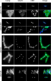
Figure 1.
Figure 1.
Formation of actomyosin rings during metaphase and anaphase in cdc15 mutant cells. cdc15-140, cdc15-287, and cdc7-24 cells were grown asynchronously at the permissive temperature and shifted to the restrictive temperature for 3 h. Spores from a cdc15Δ::ura4+/cdc15+ strain were incubated for 3 h in YES medium to assist germination, washed, and shifted to selective medium lacking uracil for 24 h. Cells were fixed and stained with α-Cdc4p antibodies, α-TAT1 antibodies, and DAPI to visualize actomyosin rings, tubulin, and DNA, respectively.
Figure 2.
Figure 2.
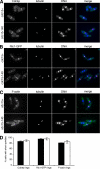
Maintenance of stable actomyosin rings in metaphase in cdc15 mutant cells. (A) pREP1-mad2+ and cdc15-140 pREP1-mad2+ cells were arrested in metaphase by growth in medium lacking thiamine for 24 h. Cells were shifted to the restrictive temperature for 3 h, fixed, and stained with α-Cdc4p antibodies, α-TAT1 antibodies, and DAPI to visualize actomyosin rings, tubulin, and DNA, respectively. (B) pREP1-mad2+ rlc1-GFP and cdc15-140 pREP1-mad2+ rlc1-GFP were grown and treated as in A except for staining with α-GFP antibodies to visualize actomyosin rings. (C) pREP1-mad2+ and cdc15-140 pREP1-mad2+ cells were grown and treated as in A except for staining with Alexa 488–conjugated phalloidin to visualize F-actin. (D) Quantitation of Cdc4p, Rlc1-GFP, and F-actin rings in cells with short (metaphase) spindles in pREP1-mad2+ [rlc1-GFP] (■) and cdc15-140 pREP1-mad2+ [rlc1-GFP] (□).
Figure 3.
Figure 3.
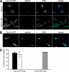
Maintenance of stable formin rings in metaphase in cdc15 mutant cells. (A) pREP1-mad2+ cdc12-GFP and cdc15-140 pREP1-mad2+ cdc12-GFP cells were arrested in metaphase by growth in medium lacking thiamine for 24 h. Cells were shifted to the restrictive temperature for 3 h, fixed, and stained with α-GFP antibodies, α-TAT1 antibodies, and DAPI to visualize formin rings, tubulin, and DNA, respectively. (B) pREP1-mad2+ cdc15-GFP cells were grown and treated as in A except for staining with α-GFP antibodies to visualize Cdc15p rings. (C) Quantitation of Cdc12-GFP rings in cells with short (metaphase) spindles in pREP1-mad2+ cdc12-GFP (■) and cdc15-140 pREP1-mad2+ cdc12-GFP (□) and of Cdc15-GFP rings in cells with short (metaphase) spindles in pREP1-mad2+ cdc15-GFP (▩).
Figure 4.
Figure 4.
Requirement of Mid1p for actomyosin ring maintenance in metaphase in the absence of functional Cdc15p. (A) mid1-18 pREP1-mad2+ rlc1-GFP and cdc15-140 mid1-18 pREP1-mad2+ rlc1-GFP cells were arrested in metaphase by growth in medium lacking thiamine for 24 h. Cells were shifted to the restrictive temperature for 3 h or kept at the permissive temperature as control, fixed and stained with α-GFP antibodies, α-TAT1 antibodies, and DAPI to visualize actomyosin rings, tubulin, and DNA, respectively. (B) Quantitation of actomyosin rings in cells with short (metaphase) spindles in mid1-18 pREP1-mad2+ rlc1-GFP at the restrictive temperature (■) and cdc15-140 mid1-18 pREP1-mad2+ rlc1-GFP at the restrictive (□; y = 0) and permissive temperature (▩).
Figure 5.
Figure 5.
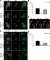
Failure to form or maintain actomyosin rings in cdc15 mutant cells when the septation initiation network is active. (A) cdc16-116 rlc1-GFP, cdc15-140 cdc16-116 rlc1-GFP and clp1Δ::ura4+ cdc16-116 rlc1-GFP cells were arrested in S phase by growth in the presence of 12 mM HU. After 6 h cells were shifted to the restrictive temperature (t = 0 min) to ectopically activate the septation initiation network. At t = 80 min, cells were fixed, stained with DAPI to visualize DNA, and examined for presence of actomyosin rings marked by Rlc1-GFP epifluorescence. (B) Quantitation of actomyosin rings in cdc16-116 rlc1-GFP cells (■), cdc15-140 cdc16-116 rlc1-GFP cells (□; y = 0.25) and clp1Δ::ura4+ cdc16-116 rlc1-GFP cells (▩). (C) cdc16-116 mid1-YFP CFP-myo2 cells were arrested in S phase as above. At t = 60 min, live cells were imaged by confocal microscopy. (D) cps1-191 rlc1-GFP and cdc15-140 cps1-191 rlc1-GFP cells were shifted to the restrictive temperature for 4 h or kept at the permissive temperature as control. Samples were fixed, stained with DAPI to visualize DNA, and examined for presence of actomyosin rings marked by Rlc1-GFP epifluorescence. (E) Quantitation of actomyosin rings in cps1-191 rlc1-GFP cells at the restrictive temperature (■) and cdc15-140 cps1-191 rlc1-GFP cells at the restrictive (□; y = 1.5) and permissive temperature (▩).
Figure 6.
Figure 6.
Cytoplasmic retention of Clp1p and spindle pole body localization of Cdc7p are maintained upon cytokinesis delay in cdc15 mutant cells. (A) cps1-191 clp1-GFP and cdc15-140 cps1-191 clp1-GFP cells were shifted to the restrictive temperature for 4 h. Samples were fixed, stained with DAPI to visualize DNA, and examined for cytoplasmic localization of Clp1-GFP (epifluorescence). (B) Quantitation of cytoplasmic Clp1-GFP localization in cps1-191 clp1-GFP cells (■) and cdc15-140 cps1-191 clp1-GFP cells (□). (C) cps1-191 cdc7-GFP and cdc15-140 cps1-191 cdc7-GFP cells were shifted to the restrictive temperature for 4 h. Samples were fixed and stained with α-GFP antibodies and DAPI to visualize Cdc7p localization and DNA, respectively. (D) Quantitation of Cdc7-GFP localization in cps1-191 cdc7-GFP cells (■) and cdc15-140 cps1-191 cdc7-GFP cells (□).
Figure 7.
Figure 7.
Hypophosphorylation of Cdc15p in at least two distinct steps, of which one is dependent on Clp1p, and maintenance of hypophosphorylation upon cytokinesis delay. (A) cdc25-22 cdc15-HA cells were arrested at the G2/M boundary by incubation at the restrictive temperature for 4 h and synchronously released by shift to the permissive temperature (t = 0 min). nda3-KM311 cdc15-HA cells were arrested at metaphase by incubation at the restrictive temperature for 6 h. Samples were taken (cdc25-22 block is t = 0 min; cdc25-22 release is t = 60 min), and total protein lysates were prepared, separated by SDS-PAGE, blotted, and detected with α-HA antibodies. Note that the protein level of Cdc15p-HA differed among the samples; total protein per lane was adjusted accordingly to compare similar amounts of Cdc15p-HA. (B) Samples from cps1-191 cdc15-HA cells and clp1Δ::ura4+ cps1-191 cdc15-HA cells incubated at the restrictive temperature for 4 h were separated by SDS-PAGE, blotted, and detected with α-HA antibodies. Membrane preparations of these samples were used because they allowed for a slightly improved resolution of Cdc15p-HA bands. (C) clp1Δ::ura4+ cdc25-22 cdc15-HA cells were arrested at the G2/M boundary by incubation at the restrictive temperature for 4 h and synchronously released by shift to the permissive temperature (t = 0 min). clp1Δ::ura4+ nda3-KM311 cdc15-HA cells were arrested at metaphase by incubation at the restrictive temperature for 6 h. These samples and samples from experiments in A (cdc25-22 block is t = 0 min; cdc25-22 release is t = 60 min for clp1+ and t = 80 min for clp1Δ) were separated by SDS-PAGE, blotted, and detected with α-HA antibodies. Note that the protein level of Cdc15p-HA differed among the samples; total protein per lane was adjusted accordingly to compare similar amounts of Cdc15p-HA.

Similar articles

Cited by

References

    1. Arai R., Mabuchi I. F-actin ring formation and the role of F-actin cables in the fission yeast Schizosaccharomyces pombe. J. Cell Sci. 2002;115:887–898. - PubMed
    1. Balasubramanian M. K., Bi E., Glotzer M. Comparative analysis of cytokinesis in budding yeast, fission yeast and animal cells. Curr. Biol. 2004;14:R806–R818. - PubMed
    1. Balasubramanian M. K., McCollum D., Chang L., Wong K. C., Naqvi N. I., He X., Sazer S., Gould K. L. Isolation and characterization of new fission yeast cytokinesis mutants. Genetics. 1998;149:1265–1275. - PMC - PubMed
    1. Balasubramanian M. K., McCollum D., Gould K. L. Cytokinesis in fission yeast Schizosaccharomyces pombe. Methods Enzymol. 1997;283:494–506. - PubMed
    1. Blondel M., Bach S., Bamps S., Dobbelaere J., Wiget P., Longaretti C., Barral Y., Meijer L., Peter M. Degradation of Hof1 by SCF(Grr1) is important for actomyosin contraction during cytokinesis in yeast. EMBO J. 2005;24:1440–1452. - PMC - PubMed

Publication types

MeSH terms

LinkOut - more resources