. 2006 Jul;17(7):3232-41.
doi: 10.1091/mbc.e05-09-0906. Epub 2006 May 10.

Aurora-B/AIM-1 regulates the dynamic behavior of HP1alpha at the G2-M transition

Affiliations

Aurora-B/AIM-1 regulates the dynamic behavior of HP1alpha at the G2-M transition

Yasuhiko Terada. Mol Biol Cell. 2006 Jul.

Abstract

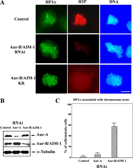
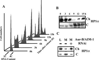
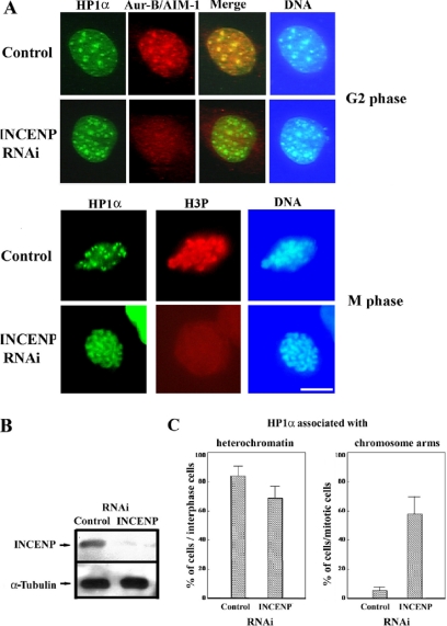
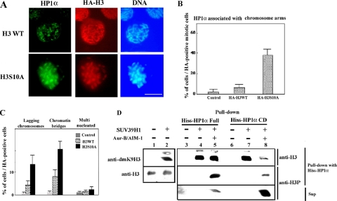
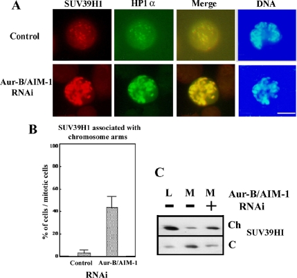
Heterochromatin protein 1 (HP1) plays an important role in heterochromatin formation and undergoes large-scale, progressive dissociation from heterochromatin in prophase cells. However, the mechanisms regulating the dynamic behavior of HP1 are poorly understood. In this study, the role of Aurora-B was investigated with respect to the dynamic behavior of HP1alpha. Mammalian Aurora-B, AIM-1, colocalizes with HP1alpha to the heterochromatin in G2. Depletion of Aurora-B/AIM-1 inhibited dissociation of HP1alpha from the chromosome arms at the G2-M transition. In addition, depletion of INCENP led to aberrant cellular localization of Aurora-B/AIM-1, but it did not affect heterochromatin targeting of HP1alpha. It was proposed in the binary switch hypothesis that phosphorylation of histone H3 at Ser-10 negatively regulates the binding of HP1alpha to the adjacent methylated Lys-9. However, Aurora-B/AIM-1-mediated phosphorylation of H3 induced dissociation of the HP1alpha chromodomain but not of the intact protein in vitro, indicating that the center and/or C-terminal domain of HP1alpha interferes with the effect of H3 phosphorylation on HP1alpha dissociation. Interestingly, Lys-9 methyltransferase SUV39H1 is abnormally localized together along the metaphase chromosome arms in Aurora-B/AIM-1-depleted cells. In conclusion, these results showed that Aurora-B/AIM-1 is necessary for regulated histone modifications involved in binding of HP1alpha by the N terminus of histone H3 during mitosis.


Figures

Figure 1.
Figure 1.
Aberrant localization of HP1α by depletion of Aurora-B/AIM-1. Immunostaining of HeLa cells expressing EGFP-HP1α with anti-H3 phos (H3P) after expression of Aurora-B/AIM-1 RNAi for 48 h. Scrambled siRNA was used as a control. After expression of Aurora-B/AIM-1 RNAi or kinase-negative Aurora-B/AIM-1 KR for 48 h, cells were swollen in a hypotonic solution, fixed in paraformaldehyde, and then permeabilized with 0.5 Triton X-100. During M phase, HP1α still remained associated with the chromosome arms in mitotic cells lacking H3 phosphorylation due to Aurora-B/AIM-1 RNAi or overexpression of kinase-negative Aurora-B/AIM-1 K106R (KR), whereas HP1α was associated with the centromeres in the control (control). Scale bar, 10 μm. (B) Immunoblotting of cell extracts of mitotic HeLa cell 48 h after expression of RNAi with different antibodies: anti-AIM-1 (Aur-B/AIM-1), Aurora-A (Aur-A), and α-Tubulin antibodies as a loading control. (C) Quantification of HP1α associated with the chromosome arms 48 h after expression of control, Aurora-A, or Aurora-B/AIM-1 RNAi. HeLa cells expressing EGFP-HP1α were synchronized, and the mitotic cells were counted and classified as indicated. Results represent the average of three independent experiments (>200 cells each); bars indicate standard deviations as described in Materials and Methods.
Figure 2.
Figure 2.
Aurora-B/AIM-1 inhibits its release of HP1α from chromatin in M-phase HeLa cells. (A) Samples of HeLa cells were synchronized, released at different time points, collected, and the DNA content analyzed by flow cytometry. (B) Synchronized cells were lysed by addition of 0.3% NP-40 in buffer N, containing 5 mM MgCl2. The Ch fraction was pelleted at 2000 × g for 5 min at 4°C in a microfuge. The C fraction was recovered. Equal amounts of proteins prepared from cytoplasmic and chromatin fractions were run on a 12% gel and blotted with anti-HP1α. (C) HeLa cells were synchronized in S phase using the double-thymidine block protocol. Mitotic cells were enriched by nocodazole treatment after release from the thymidine block. Cells were transfected with Aurora-B/AIM-1 (+) or control (−) RNAi during the second thymidine block, washed 9 h after initiation of the second thymidine block, and cultured again in the original culture medium for 4 h in the continued presence of thymidine. After removal of the drug, cells were cultured in fresh medium for 5 h and treated with nocodazole for 4–5 h. Both the Ch and C fractions were prepared from the exponentially growing cells (L) or mitotic cells (M). Immunoblots with anti-HP1α antibody was performed as shown in C.
Figure 3.
Figure 3.
Depletion of INCENP by RNAi inhibits association of Aurora-B/AIM-1 but not HP1α, with heterochromatin in the G2 phase. HeLa cells expressing EGFP-HP1α were transfected with control or INCENP siRNA. G2 phase cells were obtained by releasing cells synchronized by a double-thymidine block into fresh medium for a period of 10–12 h. M-phase cells were obtained from a thymidine/nocodazole block as shown in Figure 2C. (A) Cells treated with INCENP (INCENP RNAi) or control (RNase III siRNA) siRNAs were immunostained with anti-AIM-1 or anti-phos H3 (H3P) antibodies. DNA was stained with 50 ng/ml Hoechst 33258 to monitor the location of heterochromatin. Aurora-B/AIM-1 colocalizes with HP1α in heterochromatin. In INCENP-depleted cells, Aurora-B/AIM-1 does not associate with heterochromatin in G2 phase. During M phase, HP1α still remains associated with the chromosome arms. Bar, 10 μm. (B) For an immunoblot analysis, equal amounts of proteins prepared from HeLa cells transfected with INCENP (+) or control siRNA (−) were run on a 10% gel, blotted with anti-INCENP, and with anti-α-tubulin antibodies as a loading control. (C) Quantification of HP1α associated with interphase heterochromatin or the chromosome arms 48 h after transfection of control, or INCENP siRNAs. HeLa cells expressing EGFP-HP1α were synchronized, and the mitotic cells were counted and classified. Results are the average of three independent experiments (>200 cells each), and bars indicate standard deviations as described in Materials and Methods. Quantification of HP1α associated with the chromosome arms after expression of control, Aurora-A, or Aurora-B/AIM-1 RNAi for 48 h.
Figure 4.
Figure 4.
H3 phosphorylation is required but not sufficient to trigger dissociation of HP1α from H3 methylated at Lysine 9. (A) HA-tagged wild-type histone H3 (WT) or HA-tagged nonphosphorylatable H3 (S10A) were transfected into an EGFP-HP1α-expressing HeLa cell line. Cells were synchronized and fixed as shown in Figure 2C and visualized by GFP signal for HP1α, DAPI for DNA, and immunostaining with anti-HA (H3). Overexpression of H3 S10A, but not H3 WT, inhibited HP1α dissociation from the chromosome arms. (B) Quantification of HP1α associated with the chromosome arms 48 h after transfection of HA-H3 WT or S10A. HeLa cells expressing EGFP-HP1α were synchronized as shown in Figure 2C, stained with anti-HA antibody, and classified as indicated. The mitotic cells expressing HA-H3 were counted. Results are the average from three independent experiments (>200 cells each), and bars indicate standard deviations as described in Materials and Methods. (C) Quantitative analysis of the induction of chromosome segregation (lagging chromosomes and chromatin bridges) and cytokinesis defects (multinucleated cells) after HA-histone H3 WT or S10A mutant expression. The graph represents HA-positive cells expressing histone H3 72 h after transfection. Results are the average from three independent experiments (>200 cells each), and bars indicate standard deviations. (D) Effect of Aurora-B/AIM-1–mediated histone H3 phosphorylation on in vitro HP1α binding to histone H3 methylated at Lysine 9. Histone H3 peptide was subjected to an in vitro histone methyltransferase assay as a methyl donor. In vitro histone methyltransferase reactions were performed with human histone methyltransferase (HMTase) SUV39H1 (lanes 2, 4, 5, 7, and 8), as detected with anti-dmLys-9 H3 antibody (top, lanes 1 and 2) and H3 antibody (bottom). The methylated H3 peptides were collected and used in an His6-HP1α whole protein (lanes 3–5) or CD (lanes 6–8) pull-down assay after kinase assay in the presence or absence of FLAG-Aurora-B/AIM-1 protein. H3 phosphorylation was detected with anti-phos H3 (H3P) antibody (middle and bottom, lanes 3–8). Bound proteins were resolved by 14% SDS-PAGE and immunoblotted with anti-H3 (top).
Figure 5.
Figure 5.
Aberrant localization of SUV39H1 in mitotic chromosomes lacking Aurora-B/AIM-1. Immunofluorescence of mitotic chromosomes in control and Aurora-B/AIM-1–depleted HeLa cells stably expressing EGFP-HP1α and FLAG-tagged SUV39H1 with anti-FLAG antibody. (A) After expression of Aurora-B/AIM-1 RNAi for 48 h, cells were treated for 5 h with nocodazole, fixed, and stained with anti-FLAG antibody (SUV39HI). In cells depleted of Aurora-B/AIM-1, HP1α and SUV39H1 were localized at the metaphase chromosome arms. (B) Quantification of SUV39HI associated with the chromosome arms 48 h after transfection of control or Aurora-B/AIM-1 siRNAs. HeLa cells were synchronized. The mitotic cells were counted and classified as indicated. Results are the average of three independent experiments (>200 cells each), and bars indicate standard deviations as described in Materials and Methods. (C) HeLa cells were synchronized in S phase using the double-thymidine block protocol as shown in Figure 2C. Mitotic cells were enriched using nocodazole after release from the thymidine block. After transfection with Aurora-B/AIM-1 (+) or control (−) RNAi during the second thymidine block, HeLa cells were washed 9 h after the initiation of the second thymidine block and cultured again in the original culture medium for 4 h in the continued presence of thymidine. After removal of the drug, cells were further cultured for 5 h in fresh medium and treated with nocodazole for 4–5 h. Ch and C fractions were prepared from the exponentially growing cells (L) or mitotic cells (M). Immunoblot was performed with anti-SUV39HI antibody.

Similar articles

Cited by

References

    1. Aagaard L., Schmid M., Warburton P., Jenuwein T. Mitotic phosphorylation of SUV39H1, a novel component of active centromeres, coincides with transient accumulation at mammalian centromeres. J. Cell Sci. 2000;113:817–829. - PubMed
    1. Aasland R., Stewart A. F. The chromo shadow domain, a second chromo domain in heterochromatin-binding protein 1, HP1. Nucleic Acids Res. 1995;23:3168–3173. - PMC - PubMed
    1. Adams R. R., Carmena M., Earnshaw W. C. Chromosomal passengers and the (aurora) ABCs of mitosis. Trends Cell Biol. 2001a;11:49–54. - PubMed
    1. Adams R. R., Maiato H., Earnshaw W. C., Carmena M. Essential roles of Drosophila inner centromere protein (INCENP) and aurora B in histone H3 phosphorylation, metaphase chromosome alignment, kinetochore disjunction, and chromosome segregation. J. Cell Biol. 2001b;153:865–880. - PMC - PubMed
    1. Adams R. R., Wheatley S. P., Gouldsworthy A. M., Kandels-Lewis S. E., Carmena M., Smythe C., Gerloff D. L., Earnshaw W. C. INCENP binds the Aurora-related kinase AIRK2 and is required to target it to chromosomes, the central spindle and cleavage furrow. Curr. Biol. 2000;10:1075–1078. - PubMed

Publication types

MeSH terms