. 2006 May 10;34(9):2495-507.
doi: 10.1093/nar/gkl290. Print 2006.

The two eIF4A helicases in Trypanosoma brucei are functionally distinct

Affiliations

The two eIF4A helicases in Trypanosoma brucei are functionally distinct

Rafael Dhalia et al. Nucleic Acids Res. .

Abstract

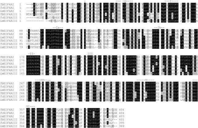
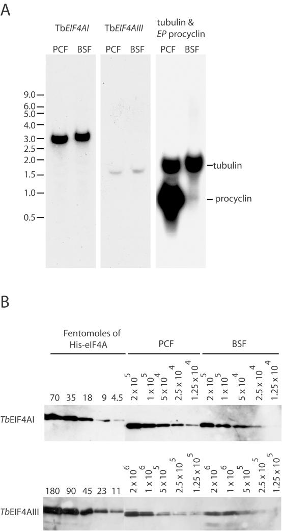
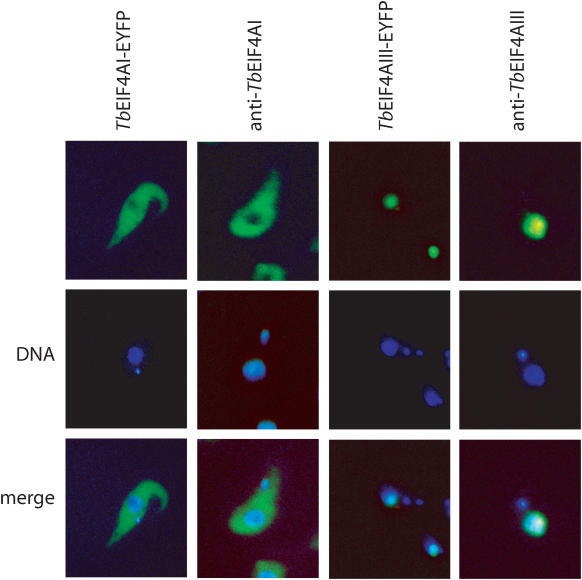
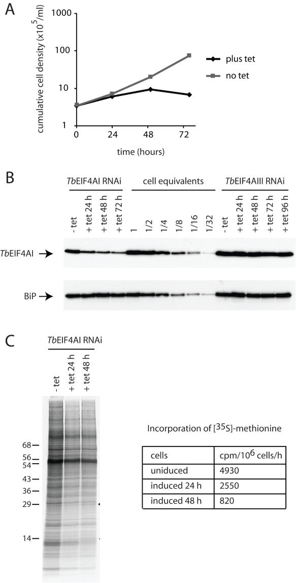
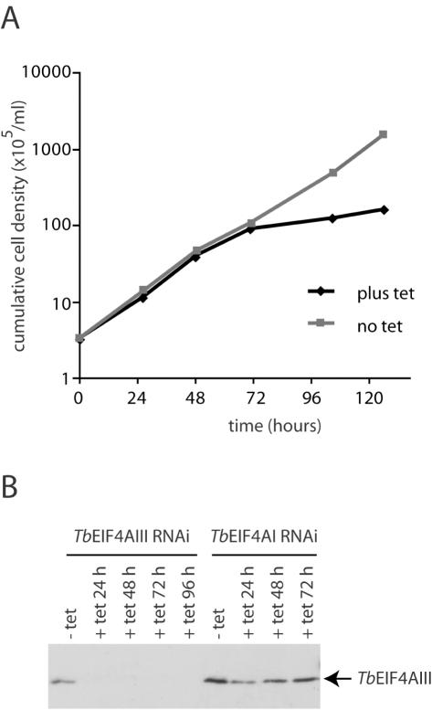
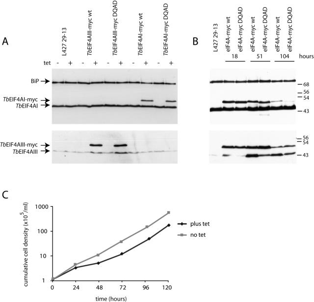
Protozoan parasites belonging to the family Trypanosomatidae are characterized by an unusual pathway for the production of mRNAs via polycistronic transcription and trans-splicing of a 5' capped mini-exon which is linked to the 3' cleavage and polyadenylation of the upstream transcript. However, little is known of the mechanism of protein synthesis in these organisms, despite their importance as agents of a number of human diseases. Here we have investigated the role of two Trypanosoma brucei homologues of the translation initiation factor eIF4A (in the light of subsequent experiments these were named as TbEIF4AI and TbEIF4AIII). eIF4A, a DEAD-box RNA helicase, is a subunit of the translation initiation complex eIF4F which binds to the cap structure of eukaryotic mRNA and recruits the small ribosomal subunit. TbEIF4AI is a very abundant predominantly cytoplasmic protein (over 1 x 10(5) molecules/cell) and depletion to approximately 10% of normal levels through RNA interference dramatically reduces protein synthesis one cell cycle following double-stranded RNA induction and stops cell proliferation. In contrast, TbEIF4AIII is a nuclear, moderately expressed protein (approximately 1-2 x 10(4) molecules/cell), and its depletion stops cellular proliferation after approximately four cell cycles. Ectopic expression of a dominant negative mutant of TbEIF4AI, but not of TbEIF4AIII, induced a slow growth phenotype in transfected cells. Overall, our results suggest that only TbEIF4AI is involved in protein synthesis while the properties and sequence of TbEIF4AIII indicate that it may be the orthologue of eIF4AIII, a component of the exon junction complex in mammalian cells.


Figures

Figure 1
Figure 1
Sequence alignment comparing the T.brucei, T.cruzi and L.major eIF4A homologues. Sequences were aligned with the Clustal W program, from the Centre for Molecular and Biomolecular Informatics (). Amino acids identical in >60% of the sequences are highlighted in dark gray, while amino acids defined as similar, based on the BLOSUM 62 Matrix, on >60% of the sequences, are shown in pale gray. When necessary, gaps were inserted within the various sequences (dashes) to allow better alignment. The nine motifs typical of DEAD-box RNA helicases (10,11) are highlighted. The single arrows indicate other individual amino acids which seems to be relevant for eIF4A function or RNA binding (12,42). Relevant GenBank accession numbers: LmEIF4AI, AAC24684/AAC24685; LmEIF4AIII, CAJ05468; TbEIF4AI, EAN76544; TbEIF4AIII, EAN79829; TcEIF4AI, EAN98527; TcEIF4AIII, EAN88971.
Figure 2
Figure 2
Expression analysis of TbEIF4AI and III. (A) Total RNA from both procyclic (PCF) and bloodstream (BSF) T.brucei forms was separated on denaturing gels and used in northern blot assays to detect the expression of TbEIF4AI and III. One of the blots was overprobed with tubulin (ubiquitously expressed) and EP procyclin (expressed in procyclics only) as controls. The migration of RNA size markers is indicated on the left in kilobases. (B) Quantification of TbEIF4AI and TbEIF4AIII in the procyclic and bloodstream forms of T.brucei. Recombinant His-tagged TbEIF4AI and III were quantified, diluted to defined concentrations (in fmol) and ran on SDS–PAGE gels with whole parasite extract obtained from known number of cells from both procyclic and bloodstream forms (1.25 × 10⁴–2 × 10⁵ for TbEIF4AI and 1.25 × 10⁵–2 × 10⁶ for TbEIF4AIII). The proteins samples were then transferred to Immobilon-P membranes followed by incubation with the affinity purified isoform specific antisera and goat anti-rabbit IgG conjugated with peroxidase, and detection by ECL. The values obtained for the abundance of the two proteins in fentomoles/105 or 106 cells were then converted in number of molecules/cell.
Figure 3
Figure 3
Subcellular localization of TbEIF4AI and III in T.brucei procyclic forms. Subcellular localization of the TbEIF4AI and III/EYFP fusion proteins in transfected T.brucei cells was examined with a fluorescence microscope. The localization of native TbEIF4AI and III was also confirmed in wild-type procyclic cells (WT 427) by indirect immunofluorescence using the TbEIF4AI or TbEIF4AIII specific antibodies followed by incubation with the fluorescein-conjugated secondary antibody. Where indicated, the cells were counterstained to locate the nuclear and kinetoplast DNA. Note lack of TbEIF4AIII staining of the kinetoplast.
Figure 4
Figure 4
RNAi of TbEIF4AI. Procyclic T.brucei cells were transfected with the p2T7-177 derived plasmid containing the TbEIF4AI gene. Transfected cells were selected after growth in the presence of phleomycin and RNA interference induced after tetracycline addition. At regular intervals, cellular growth was monitored by counting the number of viable cells, expression of TbEIF4AI assayed and total protein synthesis investigated by [35S]methionine incorporation. (A) Cell density of transfected cultures with and without tetracycline addition. (B) Western blot analysis of the time course. Note the various dilutions of total cell extract for comparison (1–1/32 cell equivalent—1 cell equivalent equals to 106 cells and was used in the various RNAi lanes). TbEIF4AI was detected with the affinity purified antisera and anti-BiP was used as a loading control. The same blot was probed with both antibodies. Equivalent extracts of cells transfected with the p2T7-177/TbEIF4AIII construct (see also Figure 5) were also used in the blot to monitor for TbEIF4AI levels. (C) [35S]methionine incorporation profile in transfected cells grown without tetracycline or 24 and 48 h after its addition. Total protein synthesis was estimated after RNAi for TbEIF4AI by incubating aliquots of the cells in the presence of [35S]methionine for 1 h followed by TCA precipitation, quantitation of the incorporated radioactivity or SDS–PAGE followed by autoradiography of the selected samples.
Figure 5
Figure 5
RNAi of TbEIF4AIII. Procyclic T.brucei cells were transfected with the p2T7-177/ TbEIF4AIII construct as described for Figure 4, monitored for cellular growth and assayed for expression of TbEIF4AIII. (A) Cell density of transfected cultures at different time points with and without tetracycline addition. (B) Western blot analysis of the time course for both the TbEIF4AIII and TbEIF4AI RNAi experiments using the TbEIF4AIII antibodies. Samples from the same experiment assayed in Figure 4B were assayed for TbEIF4AIII expression.
Figure 6
Figure 6
Expression of myc-tagged dominant negative mutants of TbEIF4AI and III in procyclic cells. (A) Western blot analysis of the expression of the various TbEIF4AI and III/myc fusions in transfected cells in the absence or after exposure to tetracycline for 18 h. In each case the expression was detected using antibodies specific to each of the eIF4A homologues. The TbEIF4AI western blot was simultaneously probed with anti-BiP as a loading control. (B) Time course expression of the different versions of TbEIF4A-myc after tetracycline addition to the culture. The TbEIF4AI western blot was simultaneously probed with anti-BiP as a loading control. (C) Effect of the expression of the dominant negative form of TbEIF4AI-myc on the growth of the transfected cells in culture.
Figure 7
Figure 7
Sequence alignment comparing TbEIF4AI and III with the putative eIF4AI and eIF4AIII from selected organisms. (A) Sequences were aligned as described in Figure 1 and the various DEAD-box motifs are shown as indicated previously. The predicted secondary structural elements derived from the modelling shown in Figure 8 and from Ref. (46) are indicated numbered α1–α13/η1–η4 (alpha-helices—H) and β1–β14 (beta-strands—S). Asterisk indicates amino acids which distinguish between the eIF4AI and eIF4AIII homologues. Further relevant GenBank accession numbers: human (Hs) eIF4AI, AAX43035; human eIF4AIII (HseIF4A3), P38919; S.pombe (Sp) eIF4A1, CAA56772; S.pombe eIF4A-like protein (Sp4Alike), CAA92238; A.thaliana (At) eIF4A1, NP_177417; A.thaliana eIF4A-like protein (At4Alike), NP_188610.
Figure 8
Figure 8
Molecular modelling of TbEIF4AI and III highlighting the position of amino acids unique to the eIF4AI or eIF4AIII homologues. Diagrams were created with the program PyMol (). (A) Ribbon diagrams of the overall structure of both TbEIF4AI and III viewed as in (46) (upper panel) or rotated 180° about its long axis (lower panel). The structure is in a closed conformation where the two, N- and C-terminal, domains are facing each other. The arrows indicate the position of the L328W substitution which lies in the loop containing Motif V and is positioned in the interface between the two domains. The dotted circles delimit the two helices discussed in the text, α5 and α10. The H/N388A/S and M213L substitutions are also indicated (their numbering differ however from the eIF4AI/eIF4AIII sequences—for instance, H388 in TbEIF4AI is equivalent to A387 in TbEIF4AIII and so on). (B) Balls and sticks representation showing the neighbourhood of the L328W substitution in both TbEIF4AI and III. The dotted lines indicate the atoms in the neighbouring amino acid chains which are positioned within a radius of 4 Å from the atoms in either the L or W residues. In both (A and B), the relevant amino acids are listed.

Similar articles

Cited by

References

    1. Campbell D.A., Thomas S., Sturm N.R. Transcription in kinetoplastid protozoa: why be normal? Microbes Infect. 2003;5:1231–1240. - PubMed
    1. Liang X.H., Haritan A., Uliel S., Michaeli S. trans and cis splicing in trypanosomatids: mechanism, factors, and regulation. Eukaryot. Cell. 2003;2:830–840. - PMC - PubMed
    1. Perry K.L., Watkins K.P., Agabian N. Trypanosome mRNAs have unusual ‘cap 4’ structures acquired by addition of a spliced leader. Proc. Natl Acad. Sci. USA. 1987;84:8190–8194. - PMC - PubMed
    1. Hershey J.W.B., Merrick W.C. Pathway and mechanism of initiation of protein synthesis. In: Sonenberg N., Hershey J.W.B., Mathews M.B., editors. Translational Control Of Gene Expression. Cold Spring Harbor, NY: Cold Spring Harbor Laboratory Press; 2000. pp. 33–88.
    1. Pestova T.V., Kolupaeva V.G., Lomakin I.B., Pilipenko E.V., Shatsky I.N., Agol V.I., Hellen C.U. Molecular mechanisms of translation initiation in eukaryotes. Proc. Natl Acad. Sci. USA. 2001;98:7029–7036. - PMC - PubMed

Publication types

MeSH terms