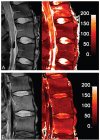
Assessment of human disc degeneration and proteoglycan content using T1rho-weighted magnetic resonance imaging
- PMID: 16688040
- PMCID: PMC2855820
- DOI: 10.1097/01.brs.0000217708.54880.51
Assessment of human disc degeneration and proteoglycan content using T1rho-weighted magnetic resonance imaging
Abstract
Study design: T1rho relaxation was quantified and correlated with intervertebral disc degeneration and proteoglycan content in cadaveric human lumbar spine tissue.
Objective: To show the use of T1rho-weighted magnetic resonance imaging (MRI) for the assessment of degeneration and proteoglycan content in the human intervertebral disc.
Summary of background data: Loss of proteoglycan in the nucleus pulposus occurs during early degeneration. Conventional MRI techniques cannot detect these early changes in the extracellular matrix content of the disc. T1rho MRI is sensitive to changes in proteoglycan content of articular cartilage and may, therefore, be sensitive to proteoglycan content in the intervertebral disc.
Methods: Intact human cadaveric lumbar spines were imaged on a clinical MR scanner. Average T1rho in the nucleus pulposus was calculated from quantitative T1rho maps. After MRI, the spines were dissected, and proteoglycan content of the nucleus pulposus was measured. Finally, the stage of degeneration was graded using conventional T2 images.
Results: T1rho decreased linearly with increasing degeneration (r = -0.76, P < 0.01) and age (r = -0.76, P < 0.01). Biochemical analysis revealed a strong linear correlation between T1rho and sulfated-glycosaminoglycan content. T1rho was moderately correlated with water content.
Conclusions: Results from this study suggest that T1rho may provide a tool for the diagnosis of early degenerative changes in the disc. T1rho-weighted MRI is a noninvasive technique that may provide higher dynamic range than T2 and does not require a high static field or exogenous contrast agents.
Figures


References
-
- Buckwalter JA. Aging and degeneration of the human intervertebral disc. Spine. 1995;20:1307–14. - PubMed
-
- Pearce RH, Grimmer BJ, Adams ME. Degeneration and the chemical composition of the human lumbar intervertebral disc. J Orthop Res. 1987;5:198–205. - PubMed
-
- Urban JP, McMullin JF. Swelling pressure of the lumbar intervertebral discs: Influence of age, spinal level, composition, and degeneration. Spine. 1988;13:179–87. - PubMed
-
- Marchand F, Ahmed AM. Investigation of the laminate structure of lumbar disc anulus fibrosus. Spine. 1990;15:402–10. - PubMed
Publication types
MeSH terms
Substances
Grants and funding
LinkOut - more resources
Full Text Sources
Other Literature Sources
Medical
Research Materials

