. 2006 May 11;441(7090):244-7.
doi: 10.1038/nature04735.

A mechanical explanation of RNA pseudoknot function in programmed ribosomal frameshifting

Affiliations

A mechanical explanation of RNA pseudoknot function in programmed ribosomal frameshifting

Olivier Namy et al. Nature. .

Abstract

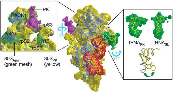
The triplet-based genetic code requires that translating ribosomes maintain the reading frame of a messenger RNA faithfully to ensure correct protein synthesis. However, in programmed -1 ribosomal frameshifting, a specific subversion of frame maintenance takes place, wherein the ribosome is forced to shift one nucleotide backwards into an overlapping reading frame and to translate an entirely new sequence of amino acids. This process is indispensable in the replication of numerous viral pathogens, including HIV and the coronavirus associated with severe acute respiratory syndrome, and is also exploited in the expression of several cellular genes. Frameshifting is promoted by an mRNA signal composed of two essential elements: a heptanucleotide 'slippery' sequence and an adjacent mRNA secondary structure, most often an mRNA pseudoknot. How these components operate together to manipulate the ribosome is unknown. Here we describe the observation of a ribosome-mRNA pseudoknot complex that is stalled in the process of -1 frameshifting. Cryoelectron microscopic imaging of purified mammalian 80S ribosomes from rabbit reticulocytes paused at a coronavirus pseudoknot reveals an intermediate of the frameshifting process. From this it can be seen how the pseudoknot interacts with the ribosome to block the mRNA entrance channel, compromising the translocation process and leading to a spring-like deformation of the P-site transfer RNA. In addition, we identify movements of the likely eukaryotic ribosomal helicase and confirm a direct interaction between the translocase eEF2 and the P-site tRNA. Together, the structural changes provide a mechanical explanation of how the pseudoknot manipulates the ribosome into a different reading frame.


Conflict of interest statement

Electron-density maps have been deposited in the European Bioinformatics Institute Electron Microscopy database, accession numbers EMD-1197, EMD-1198 and EMD-1199 (www.ebi.ac.uk/msd/iims/3D_EMdep.html). Reprints and permissions information is available at npg.nature.com/reprintsandpermissions. The authors declare no competing financial interests.

Figures

Figure 1
Figure 1. Structures of stalled 80S complexes.
a, The pseudoknot-engaged rabbit 80S ribosome (80SPK) viewed in profile. The large 60S subunit is coloured blue and the small 40S subunit is coloured yellow. The P-site tRNA stalled within the complex is coloured green, the eukaryotic translocase (eEF2) red and the pseudoknot structure (PK) purple. b, The control apo-80S ribosome (80SApo) with the subunits coloured as in b. c, The structure of a ribosome purified in the same way as 80SPK, but with the pseudoknot modified to form a stem-loop with greatly reduced capacity to induce frameshifting (80SSL). The subunits are coloured as in b and c; the P-site tRNA stalled within the complex is coloured green.
Figure 2
Figure 2. Atomic fits to the small subunit and bound cofactors of the pseudoknot-engaged ribosome.
In the centre the small subunit is viewed in a similar orientation to that of Fig. 1, with the large subunit removed computationally. The subunit, tRNA, eEF2 and pseudoknot are coloured as in Fig. 1. The yeast atomic model for the small subunit15 has been fitted to the subunit itself (blue ribbons), the structure of eEF2 (ref. 18) to the corresponding density (yellow coil), and the tRNA stalled within the complex likewise (yellow ribbon). A stereo view of the central image is provided in Supplementary Fig. 2. Left and right: close-up views, rotated as indicated to afford a detailed picture of the structural changes associated with the engaged pseudoknot (left) and the stalled tRNA (right). Left: a view from the solvent face of the 80SPK small subunit (yellow), with the 80SApo structure (green mesh) superimposed. Movements of the subunit in the 80SPK ribosome up and towards the pseudoknot structure relative to the 80SApo ribosome can be seen. Here the atomic fits (blue ribbon) are to the 80SApo structure. From this fitting the movements can be associated with the eukaryotic equivalents of the prokaryotic helicase. Although the positioning of the pseudoknot itself is clear, the mRNA passing through the entrance channel is too thin to be distinguished at the current resolution. Right: a comparison of the 80SPK (top left) and the 80SSL (top right) tRNAs (density in green; fitted atomic models in yellow). At the bottom a superposition of the two models is shown, showing the bending of the 80SPK tRNA.
Figure 3
Figure 3. A mechanical model for pseudoknot-induced -1 frameshifting.
Three different states of the small subunit translating an mRNA containing a pseudoknot that induces -1 frameshifting are shown. a, The elongating ribosome approaching the pseudoknot in the zero reading frame. b, Engagement with the pseudoknot, generating a frameshifting intermediate in which the small subunit is stalled during translocation with eEF2 bound, causing tension in the mRNA that bends the P-site tRNA in a (+ ) sense direction. As a result the anticodon–codon interaction breaks over the slippery sequence, allowing a spring-like relaxation of the tRNA in a (- ) sense direction. c, Re-engagement of the tRNA with the mRNA, leaving the ribosome translating in the -1 reading frame.

References

    1. Ogle JM, Ramakrishnan V. Structural insights into translational fidelity. Annu. Rev. Biochem. 2005;74:129–177. doi: 10.1146/annurev.biochem.74.061903.155440. - DOI - PubMed
    1. Farabaugh PJ. Programmed translational frameshifting. Microbiol. Rev. 1996;60:103–134. - PMC - PubMed
    1. Brierley, I. & Dos Ramos, F. Ribosomal frameshifting in HIV-1 and the SARS-CoV. Virus Res. advance online publication, 28 November 2005 (doi:10.1016/j.virusres.2005.10.008) - PMC - PubMed
    1. Namy O, Rousset J-P, Napthine S, Brierley I. Reprogrammed genetic decoding in cellular gene expression. Mol. Cell. 2004;13:157–168. doi: 10.1016/S1097-2765(04)00031-0. - DOI - PubMed
    1. Jacks T, Madhani HD, Masiarz FR, Varmus HE. Signals for ribosomal frameshifting in the Rous sarcoma virus gag-pol region. Cell. 1988;55:447–458. doi: 10.1016/0092-8674(88)90031-1. - DOI - PMC - PubMed

Publication types