. 2006 Jun 7;25(11):2347-57.
doi: 10.1038/sj.emboj.7601137. Epub 2006 May 11.

Cell fate-specific regulation of EGF receptor trafficking during Caenorhabditis elegans vulval development

Affiliations

Cell fate-specific regulation of EGF receptor trafficking during Caenorhabditis elegans vulval development

Attila Stetak et al. EMBO J. .

Abstract

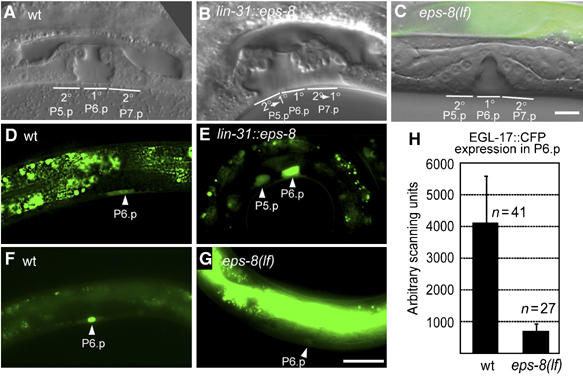
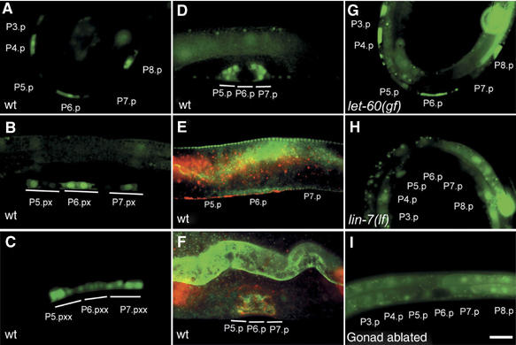
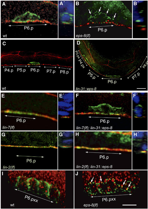
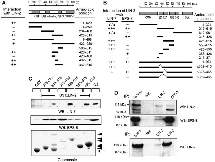
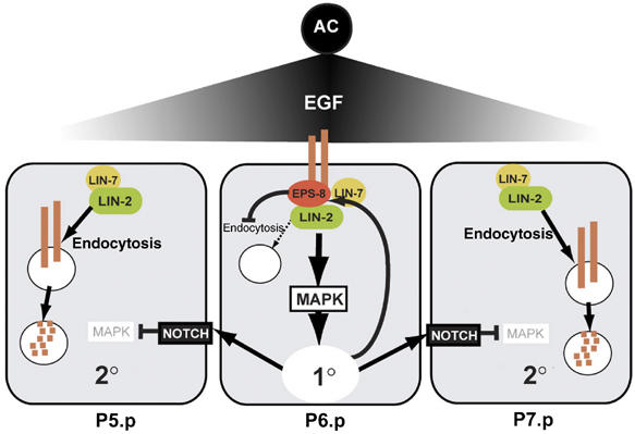
By controlling the subcellular localization of growth factor receptors, cells can modulate the activity of intracellular signal transduction pathways. During Caenorhabditis elegans vulval development, a ternary complex consisting of the LIN-7, LIN-2 and LIN-10 PDZ domain proteins localizes the epidermal growth factor receptor (EGFR) to the basolateral compartment of the vulval precursor cells (VPCs) to allow efficient receptor activation by the inductive EGF signal from the anchor cell. We have identified EGFR substrate protein-8 (EPS-8) as a novel component of the EGFR localization complex that links receptor trafficking to cell fate specification. EPS-8 expression is upregulated in the primary VPCs, where it creates a positive feedback loop in the EGFR/RAS/MAPK pathway. The membrane-associated guanylate kinase LIN-2 recruits EPS-8 into the receptor localization complex to retain the EGFR on the basolateral plasma membrane, and thus allow maximal receptor activation in the primary cell lineage. Low levels of EPS-8 in the neighboring secondary VPCs result in the rapid degradation of the EGFR, allowing these cells to adopt the secondary cell fate. Extracellular signals thus regulate EGFR trafficking in a cell type-specific manner to control pattern formation during organogenesis.


Figures

Figure 1
Figure 1
EPS-8 positively regulates vulval cell fate specification. (A) Morphology of a wild-type vulva at the L4 stage. The position of the 2° descendants of P5.p and P7.p and the 1° descendants of P6.p (out of focus) is indicated. Note that some of the 2° vulval cells remain attached to the cuticula while all 1° cells are detached. (B) Vulval morphology in a lin-31::eps-8a L4 larva showing a 2° towards 1° fate transformation of P5.p and P7.p descendants. (C) eps-8(lf) L4 larva with a wild-type vulva consisting of 22 cells. (D) Expression of the 1° cell fate marker EGL-17::GFP in a wild-type early L3 larva is restricted to P6.p. (E) Ectopic EGL-17::GFP expression in P5.p in a lin-31::eps-8a L3 larva (17% of the cases, n=24). (F) EGL-17::CFP expression in the nucleus of P6.p in a wild-type L3 larva. (G) Reduced EGL-17::CFP expression in P6.p in an eps-8(lf) L3 larva. Note in (C) and (G) the bright GFP expression in the gut due to the presence of the rescuing opt-2::eps-8::gfp transgene. Scale bar in (G) is 10 μm. (H) Quantification of EGL-17::CFP expression in P6.p of wild-type and eps-8(lf) larvae.
Figure 2
Figure 2
EPS-8 expression is upregulated in the 1° vulval cells. Expression of the eps-8p::gfp transcriptional reporter in the VPCs or their descendants of (A) a wild-type larva at the Pn.p cell stage (early L3), (B) at the Pn.px, (C) at the Pn.pxx stage (both mid L3) and (D) at the L4 stage. (E) Whole-mounts of wild-type larvae at the Pn.p cell stage and (F) at the L4 stage stained with polyclonal EPS-8 antibodies (in green). Adherens junctions are stained with MH27 in red. (G) eps-8p::gfp expression in a let-60(n1046gf) and (H) lin-7(e1413) mutant at the Pn.p stage (early L3). (I) eps-8p::gfp expression in a gonad-ablated L3 larva lacking the AC at the Pn.p stage (early L3). For a quantification of the expression patterns, see Supplementay Figure S1. Scale bar in (I) is 10 μm.
Figure 3
Figure 3
EPS-8 regulates LET-23 EGFR trafficking in the VPCs. LET-23 EGFR staining (green) in whole-mount L3 larvae using a polyclonal LET-23 antibody (see Materials and methods). The adherens junctions (red) were stained with the MH27 antibody in (A) through (H). The basal side of the VPCs is up in all panels. (A) Basolateral localization of LET-23 EGFR in P6.p of a wild-type early L3 larva. (B) Intracellular accumulation of LET-23 EGFR in an eps-8(lf) larva (arrows point at LET-23 punctae). (C) In late L2/early L3 larvae LET-23 EGFR is downregulated in all VPCs except for P6.p. (D) lin-31::eps-8a animals show persisting LET-23 EGFR expression in all VPCs. For a quantification of this phenotype, see Supplementay Figure S2. (E) Apical mislocalization of LET-23 EGFR in a lin-7(e1413) larva. (F) Partial relocalization of LET-23 EGFR to the basolateral compartment in a lin-7(e1413) larva caused by the lin-31::eps-8a transgene. (G) Apical mislocalization of LET-23 EGFR in a lin-2(n397) mutant. (H) lin-31::eps-8a fails to rescue the mislocalization of LET-23 EGFR in lin-2(n397) mutants. (I) Wild-type L3 larva (Pn.pxx stage) stained with antibodies against LET-23 (green) and the early endosomal marker EEA1 (red). (J) Partial co localization of LET-23 EGFR with EEA1 (arrows) in an eps-8(lf) larva. Panels A′, B′ and E′ to H′ show zy sections in which DAPI stained nuclei are shown in blue color. Scale bars in (D) and (J) are 5 μm.
Figure 4
Figure 4
EPS-8 associates with the LIN-2/LIN-7/LIN-10 complex. (A) The indicated fragments of C. elegans EPS-8 were tested in a yeast two-hybrid assay using amino acid 315–615 of C. elegans LIN-2 as bait. (B) The indicated fragments of C. elegans LIN-2 were tested in a yeast two-hybrid assay for interaction with full-length LIN-7 and EPS-8, respectively. Interactions were tested both by His growth and lacZ activity (+++ strong, ++ medium, + week interaction, defined by lacZ filter staining). (C) GST pull-down assays using the indicated fragments of human LIN-2 CASK cDNA fused to GST and MDCK cell lysates as source of mLIN-7A and mEPS-8 (p97 form). Bound proteins were detected on Western blots with mLIN-7A (upper panel) or mEPS-8 (middle panel) antibodies. The GST::LIN-2 fusion proteins were detected by Coomassie staining (bottom panel). Arrowheads point at GST::LIN-2 fusions, the arrow at GST. (D) Coimmunoprecipitation of mammalian LIN-2 CASK, mEPS-8 and mLIN-7A from MDCK cells. NIS indicates control precipitations with pre-immunoserum. Precipitated proteins were detected on Western blots with polyclonal LIN-2 CASK (upper panel), and monoclonal mEPS-8 antibodies (middle panel). Binding of LIN-2 CASK to mLIN-7A was detected with monoclonal LIN-2 CASK antibodies (lower panel).
Figure 5
Figure 5
The first L27 domain in LIN-2 is required for EPS-8 activity. (A) Basolateral localization of LET-23 EGFR (green) in P6.p of an early L3 lin-2(lf) larva carrying the wild-type lin-2 minigene. (B) Punctate and intracellular staining of LET-23 EGFR in an L3 larva carrying a lin-2 minigene with the Leu407 to Ser mutation in the first L27 domain. (C) Apical mislocalization of LET-23 EGFR in an L3 larva carrying a lin-2 minigene with the Ile439 to Ser mutation in the second L27 domain. Adherens junctions are stained with MH27 in red and nuclei stained with DAPI (in the z–y sections in A′, B′ and C′) are shown in blue. Scale bar in C is 10 μm. (D) Yeast two-hybrid interaction of LIN-2 carrying the indicated point mutations with LIN-7 and EPS-8, respectively, as described in Figure 4.
Figure 6
Figure 6
A model for EPS-8 function during vulval fate specification. The inductive AC signal upregulates EPS-8 expression in the 1° VPC (P6.p), where EPS-8 associates with LIN-2 to retain LET-23 EGFR on the basolateral plasma membrane. In the adjacent 2° VPCs (P5.p and P7.p) EPS-8 levels are low due to the lateral inhibition of the EGFR/RAS/MAPK signaling pathway by LIN-12 NOTCH, and LET-23 is therefore rapidly internalized and degraded in the 2° lineage.

References

    1. Berset T, Hoier EF, Battu G, Canevascini S, Hajnal A (2001) Notch inhibition of RAS signaling through MAP kinase phosphatase LIP-1 during C. elegans vulval development. Science 291: 1055–1058 - PubMed
    1. Berset TA, Hoier EF, Hajnal A (2005) The C. elegans homolog of the mammalian tumor suppressor Dep-1/Scc1 inhibits EGFR signaling to regulate binary cell fate decisions. Genes Dev 19: 1328–1340 - PMC - PubMed
    1. Brenner S (1974) The genetics of Caenorhabditis elegans. Genetics 77: 71–94 - PMC - PubMed
    1. Burdine RD, Branda CS, Stern MJ (1998) EGL-17(FGF) expression coordinates the attraction of the migrating sex myoblasts with vulval induction in C. elegans. Development 125: 1083–1093 - PubMed
    1. Burke P, Schooler K, Wiley HS (2001) Regulation of epidermal growth factor receptor signaling by endocytosis and intracellular trafficking. Mol Biol Cell 12: 1897–1910 - PMC - PubMed

Publication types

MeSH terms