

Interleukin-6 and its soluble receptor in patients with liver cirrhosis and hepatocellular carcinoma
- PMID: 16688802
- PMCID: PMC4087989
- DOI: 10.3748/wjg.v12.i16.2563
Interleukin-6 and its soluble receptor in patients with liver cirrhosis and hepatocellular carcinoma
Abstract
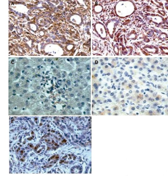
Aim: To evaluate the immunohistochemical localization of interleukin-6 (IL-6) and IL-6 receptor (IL-6R) on tumor tissue specimens from patients with hepatocellular carcinoma (HCC) and the serum levels of IL-6 and sIL-6R in a group of patients with HCC as well as liver cirrhosis (LC) in a group of patients with LC alone and in a control group.
Methods: Three groups of subjects were studied: group I (n = 83) suffering from HCC and LC, group II (n = 72) suffering from LC alone and group III (n = 42) as healthy controls. All patients had hepatitis C virus infection. Serum IL-6 and IL-6R levels were determined using a commercially available ELISA kit. Immunohistochemistry was performed using the streptavidin-biotin complex and rabbit polyclonal antibodies against IL-6 and IL-6R.
Results: Immunohistochemistry analysis showed a medium to strong cytoplasmic and membrane reactivity for IL-6 and IL-6R respectively, in at least 40% of cases of HCC, whereas liver cirrhosis patients and controls were negative for IL-6 or showed a very mild and focal dot-like cytoplasmic reaction for IL-6R. Serum IL-6 levels in HCC group were significantly higher than those in LC and control groups (P < 0.0001). There was no significant difference in sIL-6R concentrations among 3 groups. When the patients with HCC were divided into groups according to Okuda's classification, a significant serum increase of IL-6 and sIL-6R level was observed from stage I to stage III (P < 0.02, P < 0.0005). When HCC and LC patients were divided into 3 classes of cirrhosis severity according to Child-Pugh, values in HCC patients were significantly higher than those in LC patients for each corresponding class (P < 0.01).
Conclusion: IL-6 serum levels in HCC patients are higher than those in LC patients and controls, suggesting an increased production of this cytokine by neoplastic cells. sIL-6R values are similar in all groups, increasing only in stage III HCC patients. These data suggest that they have a closer relationship with the neoplastic mass rather than with the residual functioning hepatic mass.
Figures
References
-
- Oka M, Iizuka N, Yamamoto K, Gondo T, Abe T, Hazama S, Akitomi Y, Koishihara Y, Ohsugi Y, Ooba Y, et al. The influence of interleukin-6 on the growth of human esophageal cancer cell lines. J Interferon Cytokine Res. 1996;16:1001–1006. - PubMed
-
- Miki S, Iwano M, Miki Y, Yamamoto M, Tang B, Yokokawa K, Sonoda T, Hirano T, Kishimoto T. Interleukin-6 (IL-6) functions as an in vitro autocrine growth factor in renal cell carcinomas. FEBS Lett. 1989;250:607–610. - PubMed
-
- Lee JD, Sievers TM, Skotzko M, Chandler CF, Morton DL, McBride WH, Economou JS. Interleukin-6 production by human melanoma cell lines. Lymphokine Cytokine Res. 1992;11:161–166. - PubMed
-
- Siegall CB, Schwab G, Nordan RP, FitzGerald DJ, Pastan I. Expression of the interleukin 6 receptor and interleukin 6 in prostate carcinoma cells. Cancer Res. 1990;50:7786–7788. - PubMed
-
- Watson JM, Sensintaffar JL, Berek JS, Martínez-Maza O. Constitutive production of interleukin 6 by ovarian cancer cell lines and by primary ovarian tumor cultures. Cancer Res. 1990;50:6959–6965. - PubMed
Publication types
MeSH terms
Substances
LinkOut - more resources
Full Text Sources
Other Literature Sources
Medical
Research Materials
