Review
. 2006:35:277-98.
doi: 10.1146/annurev.biophys.35.040405.102126.

The ESCRT complexes: structure and mechanism of a membrane-trafficking network

Affiliations
Review

The ESCRT complexes: structure and mechanism of a membrane-trafficking network

James H Hurley et al. Annu Rev Biophys Biomol Struct. 2006.

Abstract

The ESCRT complexes and associated proteins comprise a major pathway for the lysosomal degradation of transmembrane proteins and are critical for receptor downregulation, budding of the HIV virus, and other normal and pathological cell processes. The ESCRT system is conserved from yeast to humans. The ESCRT complexes form a network that recruits monoubiquitinated proteins and drives their internalization into lumenal vesicles within a type of endosome known as a multivesicular body. The structures and interactions of many of the components have been determined over the past three years, revealing mechanisms for membrane and cargo recruitment and for complex assembly.


Figures
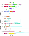
Figure 1
Figure 1
Domain structure and interactions in the ESCRT network. Protein:protein interactions within the network are indicated by solid blue lines. Interactions with lipids, ubiquitin moieties, and other proteins are shown with black arrows. For simplicity, only two of the ESCRT-III subunits, Vps20 and Snf7, are shown. The GLUE domain of human Vps36 binds to PIP3; the lipid specificity of the GLUE domain of yeast Vps36 has not been characterized.
Figure 2
Figure 2
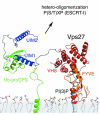
Structure of Vps27. Structures are shown where available for Vps27 proteins (FYVE (69), UIM1-ubiquitin complex (103), UIM1-UIM2 (103)), otherwise modeled on the basis of the closely related structure of the Hrs-VHS domain (64). Linker regions between the domains were generated arbitrarily and are shown only to indicate the length of the segment. The C-terminal putative Hse1-binding domain and the extended region of Vps27 are not shown. Ubiquitin is shown fused via an isopeptide bond between Gly-76 and Lys-8 of the prototypical cargo pro-carboxypeptidase S, and the transmembrane helix of pro-CPS was modeled as an ideal helix. Membrane docking of Vps27 is based on the computationally predicted optimal docking mode for the FYVE domain.
Figure 3
Figure 3
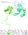
Structure of ESCRT-I. Structures are shown for the UEV domain of human Vps23 in complex with ubiquitin (102) and the HIV-1 p6 PTAP-containing peptide (82). The remainder of the ESCRT-I structure is not yet available.
Figure 4
Figure 4
Structure of ESCRT-II. The structure of the ESCRT-II core (42) is shown docked to a membrane on the basis the interaction between its Vps25 subunit and the membrane-bound ESCRT-III subunit Vps20 (104) (myristoyl group on Vps20 not shown for simplicity). The uncomplexed NZF1 domain and the NZF2-ubiquitin complex are modeled on the basis of the Npl4-NZF structure and ubiquitin complex (1). The GLUE domain (a variant of the GRAM domain, which is in turn a variant of the PH domain) is modeled and docked to the membrane on the basis of the GRAM domain of the lipid phosphatase MTMR2 (11). The GLUE domain of human Vps36 binds to PI(3,4,5)P3 and ubiquitin at sites that have yet to be determined (99), and are not shown. The binding properties of the yeast Vps36 GLUE domain have yet to be reported. The dashed line between the GLUE domain and Vps36 core winged helix (WH) region indicates residues 290-395 of Vps36, whose structure is unknown.
Figure 5
Figure 5
Structure of ESCRT-III. The structure of ESCRT-III is unknown. This cartoon shows a tripartite variant of the bipartite model proposed by Hanson and colleagues (62). The C-terminal anionic regions of ESCRT-III subunits is roughly twice as large as the N-terminal basic region, and appears to have functions both in oligomerization of ESCRT-III on membranes and in binding to other proteins, such as Vps4. Black arrows indicate directions of in which the oligomeric array can grow.
Figure 6
Figure 6
Structure of Bro1. The structure of the Bro1 domain (56), which comprises the N-terminal half of Bro1, is shown docked to a negatively curved membrane. The interaction of this domain with negatively curved membranes is suggested by the shape of the structure and by the properties of the full-length human Bro1 homologue; however, this interaction and docking mode are speculative and have yet to be directly tested.
Figure 7
Figure 7
Structure of Vps4. The modeled structure of the Vps4 AAA domain hexamer was modeled (93) is shown together with the corresponding six copies of the MIT domain (94). For simplicity, a single subunit of the ESCRT-III complex is shown engaged to a single MIT domain. The ESCRT-III subunit is thought to be fed through the central pore in the hexamer (not shown in this view) as ATP is hydrolyzed by the AAA domain.
Figure 8
Figure 8
The ESCRT complexes in MVB Sorting - The Vps27 protein complex initiates the MVB sorting process. It is targeted to endosomal membranes via its FYVE domain that binds PI(3)P, and its UIM domains which bind ubiquitinated MVB cargo such as carboxypeptidase S (CPS). Vps27 subsequently recruits and activates the ESCRT-I complex via the P(S/T)XP motif in the C-terminal domain of Vps27 that interacts with the UEV domain of Vps23 in ESCRT-I. Ubiquitinated cargo is recognized by ESCRT-I (via the UEV domain of Vps23) and by ESCRT-II (via the NZF domain in Vps36). ESCRT-III is required for concentration of cargoes into MVB vesicles and coordinates the association of accessory factors such as Bro1 and the Doa4 deubiquitinating enzyme that removes ubiquitin from cargo. The AAA-type ATPase Vps4 plays a critical role in catalyzing the dissociation of the ESCRT complexes. Together, these proteins appear to direct MVB vesicle formation, cargo sorting into MVB vesicles and vesicle fission. See text for further details.

References

    1. Alam SL, Sun J, Payne M, Welch BD, Black BK, et al. Ubiquitin interactions of NZF zinc fingers. EMBO J. 2004;23:1411. - PMC - PubMed
    1. Amerik AY, Li SJ, Hochstrasser M. Analysis of the deubiquitinating enzymes of the yeast Saccharomyces cerevisiae. Biol. Chem. 2000;381:981. - PubMed
    1. Amit I, Yakir L, Katz M, Zwang Y, Marmor MD, et al. Tal, a Tsg101- specific E3 ubiquitin ligase, regulates receptor endocytosis and retrovirus budding. Genes Dev. 2004;18:1737. - PMC - PubMed
    1. Antonyak MA, McNeill CJ, Wakshlag JJ, Boehm JE, Cerione RA. Activation of the Ras-ERK pathway inhibits retinoic acid-induced stimulation of tissue transglutaminase expression in NIH3T3 cells. J. Biol. Chem. 2003;278:15859. - PubMed
    1. Asao H, Sasaki Y, Arita T, Tanaka N, Endo K, et al. Hrs is associated with STAM, a signal-transducing adaptor molecule - Its suppressive effect on cytokine-induced cell growth. J. Biol. Chem. 1997;272:32785. - PubMed

Publication types

MeSH terms

LinkOut - more resources