. 2006 Aug 15;398(1):23-36.
doi: 10.1042/BJ20060423.

Scanning peptide array analyses identify overlapping binding sites for the signalling scaffold proteins, beta-arrestin and RACK1, in cAMP-specific phosphodiesterase PDE4D5

Affiliations

Scanning peptide array analyses identify overlapping binding sites for the signalling scaffold proteins, beta-arrestin and RACK1, in cAMP-specific phosphodiesterase PDE4D5

Graeme B Bolger et al. Biochem J. .

Abstract

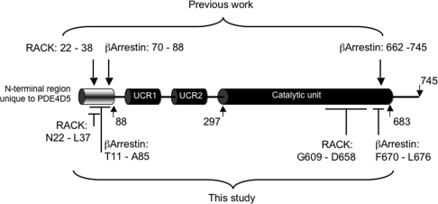
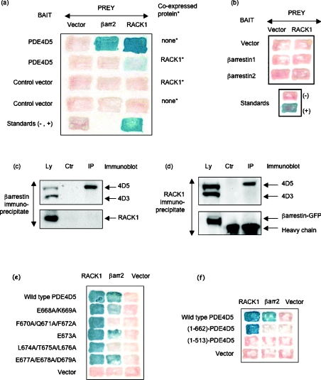
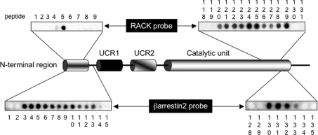
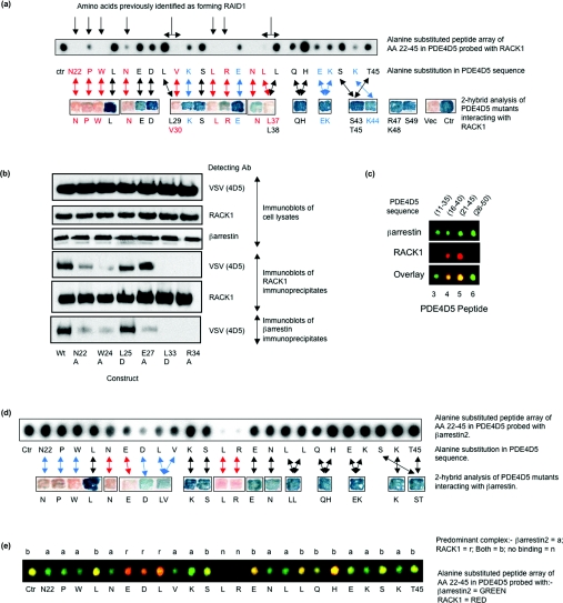
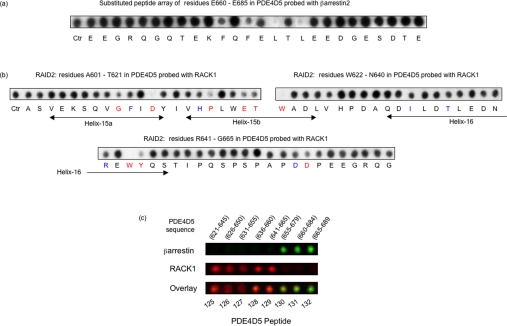
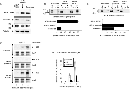
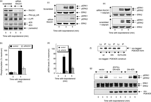
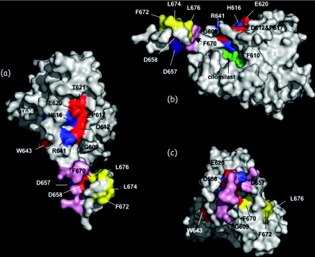
The cAMP-specific phosphodiesterase PDE4D5 can interact with the signalling scaffold proteins RACK (receptors for activated C-kinase) 1 and beta-arrestin. Two-hybrid and co-immunoprecipitation analyses showed that RACK1 and beta-arrestin interact with PDE4D5 in a mutually exclusive manner. Overlay studies with PDE4D5 scanning peptide array libraries showed that RACK1 and beta-arrestin interact at overlapping sites within the unique N-terminal region of PDE4D5 and at distinct sites within the conserved PDE4 catalytic domain. Screening scanning alanine substitution peptide arrays, coupled with mutagenesis and truncation studies, allowed definition of RACK1 and beta-arrestin interaction sites. Modelled on the PDE4D catalytic domain, these form distinct well-defined surface-exposed patches on helices-15-16, for RACK1, and helix-17 for beta-arrestin. siRNA (small interfering RNA)-mediated knockdown of RACK1 in HEK-293 (human embryonic kidney) B2 cells increased beta-arrestin-scaffolded PDE4D5 approx. 5-fold, increased PDE4D5 recruited to the beta2AR (beta2-adrenergic receptor) upon isoproterenol challenge approx. 4-fold and severely attenuated (approx. 4-5 fold) both isoproterenol-stimulated PKA (protein kinase A) phosphorylation of the beta2AR and activation of ERK (extracellular-signal-regulated kinase). The ability of a catalytically inactive form of PDE4D5 to exert a dominant negative effect in amplifying isoproterenol-stimulated ERK activation was ablated by a mutation that blocked the interaction of PDE4D5 with beta-arrestin. In the present study, we show that the signalling scaffold proteins RACK1 and beta-arrestin compete to sequester distinct 'pools' of PDE4D5. In this fashion, alterations in the level of RACK1 expression may act to modulate signal transduction mediated by the beta2AR.


Figures

Figure 1
Figure 1. Location of sites for RACK1 and β-arrestin interaction on PDE4D5
A schematic of PDE4D5 with its unique N-terminal region, upstream conserved region 1 (UCR1), upstream conserved region 2 (UCR2), catalytic region and extreme C-terminal region. Indicated above the schematic are the sites of β-arrestin and RACK1 interaction identified prior to the present study. Below it are interaction sites arising from the present study.
Figure 2
Figure 2. RACK1 and β-arrestin interact with PDE4D5 in a mutually exclusive fashion
(a) Two-hybrid analysis with PDE4D5 as ‘bait’ and either β-arrestin 2 (βarr2) or RACK1 as ‘prey’. Where indicated, RACK1 was additionally co-expressed as a ‘competitor’. Blue indicates a positive interaction and pink a null interaction. Controls are vectors without inserts. (b) A two-hybrid assay was performed, as in (a). Also shown is the positive standard and negative (‘empty’ vector) control for this experiment (‘standards’), which are both identical with those in (a). These data are typical of results obtained at least three times. (c) Immunoblots of endogenous PDE4D3, PDE4D5 and RACK1 from HEK-293B2 cell lysates (Ly) and β-arrestin immunoprecipitates (IP). (d) Using a RACK1-specific antibody, endogenous RACK1 was immunoprecipitated (IP) from HEK-293B2 cells transfected to express β-arrestin–GFP. Lysates (Ly) and RACK1 immunoprecipitates (IP) were immunoblotted for endogenous PDE4D3, PDE4D5 and transfected β-arrestin–GFP. No immunoreactive species were detected in control (Ctr) experiments using pre-immune antisera. These data are typical of experiments performed at least three times. (e) Various mutations in the EKFQFELTLEE motif of PDE4D5 (amino acids 668–678) were tested in two-hybrid assays for their ability to block the interaction of PDE4D5 with RACK1 and β-arrestin 2. A two-hybrid assay was performed, as in (a). Standards (not shown) were identical with those in (a). (f) Various C-terminal deletions of PDE4D5 were tested for their ability to interact with either RACK1 or β-arrestin 2. A two-hybrid assay was performed, as in (a). Standards (not shown) were identical to those in (a).
Figure 3
Figure 3. Probing PDE4D5 peptide arrays for RACK1 and β-arrestin interaction sites
PDE4D5 is shown schematically as in Figure 1. Data show immobilized peptide ‘spots’ of overlapping 25-mer peptides each shifted along by five amino acids in the entire PDE4D5 sequence probed for interaction with either β-arrestin 2–GST or RACK1–GST and detection by immunoblotting. Positively interacting peptides generate dark spots whereas non-interacting peptide leave white (blank) spots. In all other sections of the array, spots were blank with either probe. Spot numbers relate to peptides in the scanned array and whose sequence is given in Table 1. The disposition of the peptides in PDE4D5 sequence are shown relative to identified domains in a schematic of PDE4D5, which indicates its isoform-specific N-terminal region, upstream conserved regions 1 and 2 (UCR1, UCR2), catalytic unit and PDE4D C-terminal region.
Figure 4
Figure 4. Binding of RACK1 and β-arrestin to sequential alanine-substituted versions of a RAID1-containing peptide
(a) The row of spots in the upper panel shows a peptide array based upon a 25 mer ‘parent’ peptide of sequence Asn22–Thr45 in PDE4D5. Ctr refers to the unmutated peptide and all other spots reflect peptide ‘progeny’ where indicated amino acids were sequentially and individually substituted for alanine. This array was probed with RACK1–GST and interaction was determined by immunoblotting with an anti-RACK1 antibody. The lower panel of coloured patches shows various mutant forms of PDE4D5, with the indicated amino acids substituted for alanine, probed for interaction with RACK1 in a two-hybrid assay (positive, blue; and null, pink). For substitution/mutation at specific residues, then red indicates abolition of binding using both assay procedures, whereas black arrows indicate binding is still apparent. Blue arrows indicate differences between the two assay procedures. (b) HEK-293B2 cells were transfected with either wild-type or the indicated mutant forms of VSV-epitope-tagged PDE4D5. Lysates and immunoprecipitates were immunoblotted as indicated. As β-arrestin migrates on SDS/PAGE identically with Ig heavy chain, we were unable to immunoblot for β-arrestin in immunoprecipitates. Equal protein loading was used in each track. Lysates contained 1/20 protein loading compared with the immunoprecipitates. (c) Odyssey® analysis of PDE4D5 peptides 3–6 (see Figure 3) challenged with equimolar (200 nM) β-arrestin 2–GST and RACK1–GST. β-Arrestin 2 (green) and RACK1 (red) binding were detected simultaneously, with dual binding (yellow) shown in the combined channels (Overlay). Null binding is black. Data are typical of those obtained using three separately synthesized arrays. (d) Peptide array and two-hybrid assays performed as in (a), but β-arrestin 2 was used in the two screens instead of RACK1. (e) Performed as in (c) to probe β-arrestin 2–GST and RACK1–GST binding simultaneously, but with the alanine scanning peptide arrays used in both (a) and (d). Data are typical of experiments performed at least three times. AA, amino acid.
Figure 5
Figure 5. Defining the binding of RACK1 and β-arrestin to the PDE4D catalytic unit using scanning peptide arrays
(a) Scanning Glu660–Glu685 in PDE4D5 for interaction with β-arrestin–GST. Anti-(β-arrestin) antisera was used for detection; all other conditions were as in Figure 4(a). Native peptide (Ctr) plus progeny with the indicated residue substituted for alanine. (b) Scanning of Ala601–Glu665 in PDE4D5 for interaction with RACK1–GST. Three peptides were used to cover this region (Ctr) with conditions as in Figure 4(a). The indicated residues were substituted for alanine, unless the indicated amino acid was alanine in native PDE4D5, when it was substituted for aspartate. Mutation of amino acids coloured red indicate null interaction (<10%), whereas those coloured blue indicate reductions >50% based upon densitometric analysis of spots for n=3 experiments. (c) Odyssey® analysis of PDE4D5 peptides 125–132 (see Figure 3) incubated with equimolar (200 nM) β-arrestin 2–GST and RACK1–GST. Detection was as in Figure 4(c). Data are typical of experiments performed at least three times.
Figure 6
Figure 6. siRNA-mediated knockdown of RACK1 and β-arrestin on PDE4D5 recruitment to the β2AR
(a) HEK-293B2 cells were subject to siRNA knockdown of either β-arrestin, RACK1 or with scrambled siRNA. Lysates were immunoblotted for endogenous RACK1, β-arrestin, tubulin, PDE4D3 and PDE4D5, as indicated. (b) Cells were treated with siRNA as above and immunoprecipitates generated from lysates (Ly) using either a β-arrestin-specific antiserum (IP) or with non-specific serum (Ctr). Note that loading blots cannot be performed for β-arrestin as it co-migrates with Ig heavy-chain. Upper panel shows immunoblots, and lower panel shows a quantitative analysis of three independent experiments. (c) As in (b), except that RACK1 immunoprecipitates were probed for PDE4D. (d) Cells were treated with siRNA as in (a) and then treated with isoproterenol (10 μM) for the indicated times. The β2AR was selectively immunoprecipitated (IP) and probed for PDE4D5. (e) A quantitative analysis of three independent experiments performed as in (d). Data are means±S.D.
Figure 7
Figure 7. siRNA-mediated knockdown of RACK1 on β2AR phosphorylation by PKA and activation of ERK in HEK-293B2 cells
(a) Cells were transfected with either scrambled siRNA or siRNA specific for RACK1 prior to challenge with isoproterenol (10 μM for 5 min). Equal amounts of cell lysate were immunoblotted for RACK1, total β2AR, PKA-phosphorylated form of the β2AR, β-arrestin and PDE4D. (b) Quantification of the PKA phosphorylation status of the β2AR upon isoproterenol (10 μM) challenge (5 min) of cells after prior transfection with either scrambled siRNA or siRNA specific for RACK1 in three separate experiments performed as in (a). (c) As in (a), but lysates were immunoblotted for total ERK and phospho-ERK. (d) Quantification of ERK phosphorylation (n=3). (e) Cells treated with either RACK1 siRNA or scrambled siRNA with subsequent challenge using isoproterenol plus the PDE4 selective inhibitor, rolipram (10 μM). Lysates were immunoblotted for total ERK and phospho-ERK (pERK). (f) Cells were transfected with VSV-epitope-tagged versions of wild-type PDE4D5 (wt), D556A-PDE4D5 (DN) or E26A:D556A-PDE4D5 (E27A-DN). β-Arrestin immunoprecipitates (IP) were generated from cell lysates (Ly) and both were immunoblotted with a VSV-specific antibody to detect recombinant PDE4D5. (g) Cells were transfected as (f) and then challenged for the indicated times with isoproterenol (10 μM). Equal amounts of lysate were immunoblotted to identify VSV-tagged PDE4D, total ERK and phosphorylated ERK. Data are typical of experiments performed at least three times. Data are means±S.D.
Figure 8
Figure 8. Structural representation of binding sites for RACK1 and β-arrestin on the PDE4D structural models
(a and b) Two different perspectives of the structural model of the 324–677 stretch of the PDE4D generated, as described in the Experimental section, using 1F0J PDE4B structure as a template. Amino acids whose substitution with alanine leads to loss of RACK1 interaction signal are shown in red, and those where substitution leads to a clear reduction in interaction signal are shown in blue. Amino acids whose substitution with alanine leads to loss of β-arrestin interaction signal are shown in yellow, the flexible linker between helices- 16 and 17 is shown in pink, and the 1XOM inhibitor cilomilast is shown in spacefill representation in green. (c) The structural model of the 324–677 stretch of the PDE4D generated using 1XM6 PDE4B structure as a template. Colour-coding as above. These structural Figures were created using the PyMOL program (http://pymol.sourceforge.net/).

Similar articles

Cited by

References

    1. Hall R. A. β-Adrenergic receptors and their interacting proteins. Semin. Cell. Dev. Biol. 2004;15:281–288. - PubMed
    1. Lefkowitz R. J., Shenoy S. K. Transduction of receptor signals by β-arrestins. Science. 2005;308:512–517. - PubMed
    1. Wong W., Scott J. D. AKAP signalling complexes: focal points in space and time. Nat. Rev. Mol. Cell. Biol. 2004;5:959–970. - PubMed
    1. Penela P., Ribas C., Mayor F., Jr Mechanisms of regulation of the expression and function of G protein-coupled receptor kinases. Cell. Signalling. 2003;15:973–981. - PubMed
    1. Baillie G. S., Houslay M. D. Arrestin times for compartmentalised cAMP signalling and phosphodiesterase-4 enzymes. Curr. Opin. Cell. Biol. 2005;17:129–134. - PubMed

Publication types

MeSH terms