. 2006 Sep 1;398(2):233-42.
doi: 10.1042/BJ20060004.

Novel genes encoding six kinds of three-finger toxins in Ophiophagus hannah (king cobra) and function characterization of two recombinant long-chain neurotoxins

Affiliations

Novel genes encoding six kinds of three-finger toxins in Ophiophagus hannah (king cobra) and function characterization of two recombinant long-chain neurotoxins

Jing Li et al. Biochem J. .

Abstract

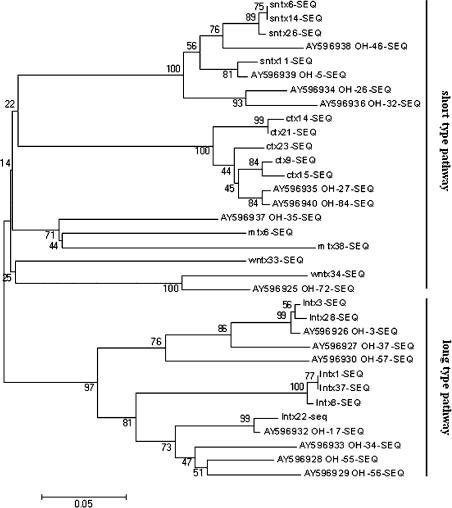
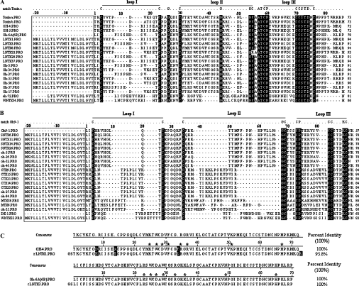
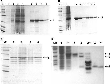
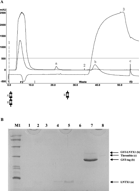
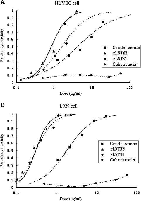
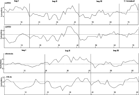
Three-finger toxins are a family of low-molecular-mass toxins (<10 kDa) having very similar three-dimensional structures. In the present study, 19 novel cDNAs coding three-finger toxins were cloned from the venom gland of Ophiophagus hannah (king cobra). Alignment analysis showed that the putative peptides could be divided into six kinds of three-finger toxins: LNTXs (long-chain neurotoxins), short-chain neurotoxins, cardiotoxins (CTXs), weak neurotoxins, muscarinic toxins and a toxin with a free SH group. Furthermore, a phylogenetic tree was established on the basis of the toxin cDNAs and the previously reported similar nucleotide sequences from the same source venom. It indicated that three-finger-toxin genes in O. hannah diverged early in the course of evolution by long- and short-type pathways. Two LNTXs, namely rLNTX1 (recombinant LNTX1) and rLNTX3, were expressed and showed cytolytic activity in addition to their neurotoxic function. By comparing the functional residues, we offer some possible explanations for the differences in their neurotoxic function. Moreover, a plausible elucidation of the additonal cytolytic activity was achieved by hydropathy-profile analysis. This, to our knowledge, is the first observation that recombinant long chain alpha-neurotoxins have a CTX-like cytolytic activity.


Figures

Figure 1
Figure 1. Phylogenetic analysis of O. hannah cDNAs obtained in the present study and of already registered cDNAs
Multiple sequences were aligned using CLUSTALW (version 1.83). The tree was reconstructed using distance methods of Neighbour-joining on the basis of the Poisson-corrected amino acid distance. The reliability of clusters was estimated using a Bootstrap test. The GenBank® accession numbers of cDNAs whose sequences have already been published are indicated. The scale bar means 0.05 amino acid substitution per site.
Figure 2
Figure 2. Multiple sequence alignments of deduced amino acids with those of toxins from O. hannah
Natural peptides for which sequences have already been published were Toxin a (Swiss-Prot. number P01387), Toxin b (P01386), OH-4 (P80516), OH-5 (P80965), Oh-6A/6B (P82662), Oh9-1 (P83302) and DE-1 (P01412). The sequences of Oh-3, -5, -17, -26, -27, -32, -34, -35, -37, -46, -55, -56, -57 and -84 were deduced from their corresponding cDNAs with GenBank® accession numbers from AY596925 to AY596940 [23]. Signal peptides are boxed. Amino acid residues that matched the template residues in the first line of each group reversed-out (i.e. white-on-black) in (A) and (B), whereas residues reversed-out in (C) are unmatched ones. The last number in each row indicates the numbers of residues in each toxin. (A) Group A toxins having ten conserved cysteine residues (five disulphide bridges). (B) Group B toxins having eight conserved cysteine residues (four disulphide bridges). (C) Alignments of rLNTX1 and rLNTX3 to their corresponding natural proteins. Functional residues at homologous sites are indicated with asterisks (*).
Figure 3
Figure 3. SDS/PAGE analysis of rLNTX3 and rLNTX1
(A)–(C) employed Tris/glycine/SDS/16%-(w/v)-PAGE and (D) employed Tris/tricine/SDS/16%-PAGE. Lane M1, protein markers of low molecular mass (kDa): 97, 66, 43, 31, 20 and 14; lane M2, peptide markers (kDa): 16, 14, 10, 0.8, 0.6 and 0.2; bands labelled 1, 2 and 3 are fusion protein, GST-tag and recombinant peptides respectively. (A) Total proteins of uninduced and induced cultures together with GST–LNTX3 obtained from each of the purification steps. Lanes 1 and 2, total proteins of culture at 37 °C after 0 and 4 h induction respectively; lanes 3 and 4, total proteins of culture at 16 °C after 0 and 20 h induction respectively; lanes 5 and 6, elution fractions from the affinity column; lanes 7 and 8, purified GST–LNTX3 from the Superdex75 column. (B) Total proteins of uninduced and induced culture together with GST–LNTX1 obtained from each of the purification steps. Lanes 1 and 2, total proteins of culture at 16 °C after 0 and 20 h induction respectively; lanes 3–5, elution fractions from the affinity column; lanes 7 and 8, purified GST–LNTX1 from the Superdex75 column. (C) Proteins from supernatant and pellet of lysate after the induction of IPTG for 20 h at 16 °C. Lanes 1 and 2, the pellet and supernatant of LNTX1 respectively; lanes 3 and 4, the pellet and supernatant of LNTX3 respectively. (D) Cleavage of fusion protein and purified recombinant peptides. Lanes 1 and 2, total proteins of uninduced and induced culture of LNTX1; lanes 3 and 4, GST–LNTX1 after being mixed with thrombin for 0 and 8 h; lanes 6 and 7, the purified rLNTX1 and rLNTX3 respectively.
Figure 4
Figure 4. Purification of recombinant peptides on a benzamidine FF column placed in series below the GSTrap FF column
(A) Chromatography of rLNTX1. Position 1 indicates the sample application. Positions 2 and 3 show the beginning of the high-salt elution and low-pH-buffer elution respectively. The high-salt-wash buffer contained 20 mM sodium phosphate and 1.0 M NaCl, pH 7.0; the low-pH-wash buffer contained 10 mM HCl and 0.5 M NaCl, pH2.0. Column A was a GSTrap FF 1-ml-volume column; column B was a HiTrap Benzamidine FF 1 ml column; ‘a’ is the released rLNTX1 peptide; ‘b’ is a mixture of GST-tag and some non-cleaved fusion proteins; ‘c’ is bovine thrombin. Abbreviation: mAU, milli-absorbance unit. (B) Tris/glycine/SDS/16%-PAGE analysis of each peak in the above chromatography. Lane M1, protein markers of low molecular mass; lanes 1–3, flow-through during sample loading; lanes 4–6, peak ‘a’ in (A); lane 7, peak ‘b’ in (A); lane 8, A chain (31 kDa) of bovine thrombin (‘c’ in A).
Figure 5
Figure 5. Characterizations of rLNTX1 and rLNTX3
(A) and (B) MALDI–TOF-MS of rLNTX1 and rLNTX3, respectively. (C) and (D) CD spectra showing the molecular ellipticity (Mol. Ellip.) of rLNTX1 and rLNTX3 respectively.
Figure 6
Figure 6. Inhibitory effects of rLNTX1 and rLNTX3 on nAChR-enriched skeletal myocytes
Cells were held throughout the experiment at −70 mV. (A) Reproducible control currents excited by 80 μM nicotine. (B) Currents inhibited by rLNTX3 at a concentration of: 1, 0.159 μM; 2, 0.318 μM; 3, 0.636 μM; 4,1.59 μM; and 5, 3.18 μM. (C) Currents inhibited by rLNTX1 at a concentration of: 1, 0.20 μM; 2, 0.50 μM; 3, 1.00 μM; 4, 5.00 μM; and 5, 10.00 μM. (D) Analysis of the data by the median-effect plot. The median effect concentrations (IC50) of each peptide are marked. (E) Fraction of inhibited current to control current (fa) as functions of recombinant peptides at a series of concentrations (labelled beside the data points).
Figure 7
Figure 7. Lethal effect of crude venom, rLNTX1, rLNTX3 and Cbtx on HUVEC and L929 cells
Figure 8
Figure 8. Hydropathy profiles for rLNTX1, rLNTX3, Cbtx and CTX-4b
Hydropathy profiles were analysed according to the Kyte–Doolittle method [35]. Values above the axis denote hydrophilic regions, whereas those below the axis indicate hydrophobic regions. Analyses were carried out using ANTHEPROT V5.0 (Institut de Biologie et Chimie des Protéines, Lyon, France).

Similar articles

Cited by

References

    1. Ricciardi A., le Du M.-H., Khayati M., Dajas F., Jean-Claude B., Menez A., Ducancel F. Do structural deviations between toxins adopting the same fold reflect functional differences? J. Biol. Chem. 2000;275:18302–18310. - PubMed
    1. Hall Z. W. α-Neurotoxins and their relatives: foes and friends? Neuron. 1999;23:4–5. - PubMed
    1. Jerusalinsky D., Harvey A. L. Toxins from mamba venoms: small proteins with selectivities for different subtypes of muscarinic acetylcholine receptors. Trends Pharmacol. Sci. 1994;15:424–430. - PubMed
    1. Utkin Y. N., Kukhtina V. V., Kryukva E. V., Chiodini F., Bertrand D., Methfessel C., Tsetlin V. I. “Weak toxin” from Naja kaouthia is a nontoxic antagonist of α7 and muscle-type nicotinic acetylcholine receptors. J. Biol. Chem. 2001;276:15810–15815. - PubMed
    1. Harvey A. L. Cardiotoxins from cobra venoms: possible mechanism of action. J. Toxicol. Toxin Rev. 1985;4:41–69.

MeSH terms