. 2006 Jul 15;574(Pt 2):457-76.
doi: 10.1113/jphysiol.2006.109116. Epub 2006 May 11.

Short-term reorganization of input-deprived motor vibrissae representation following motor disconnection in adult rats

Affiliations

Short-term reorganization of input-deprived motor vibrissae representation following motor disconnection in adult rats

Gianfranco Franchi et al. J Physiol. .

Abstract

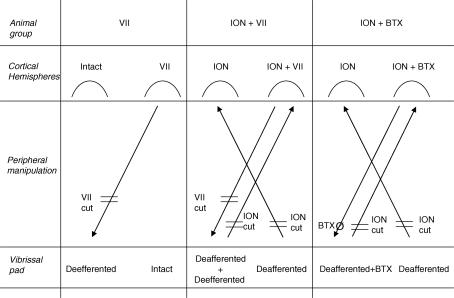
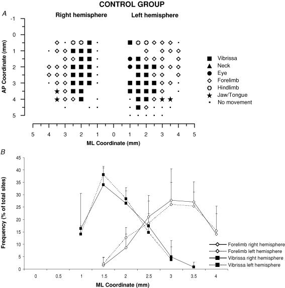
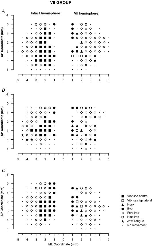
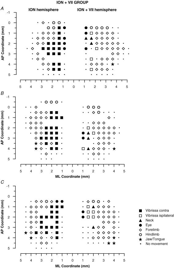
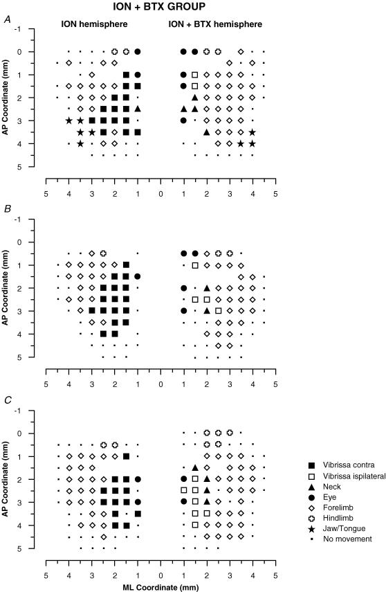
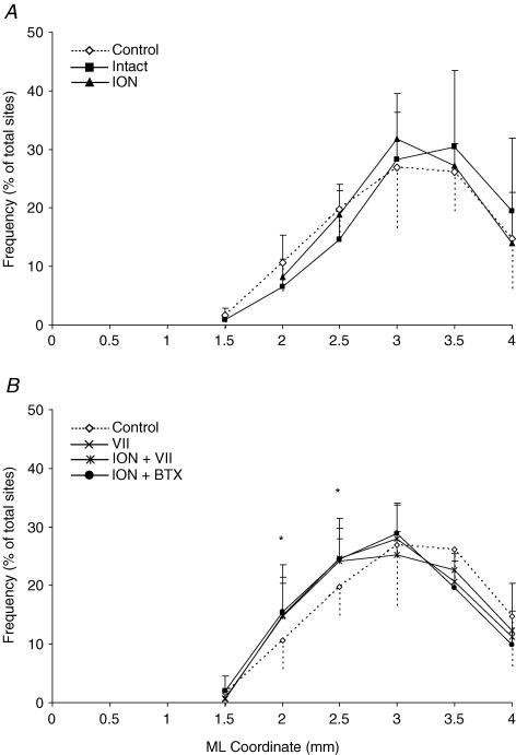
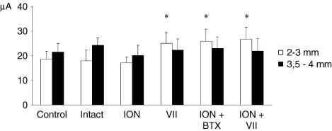
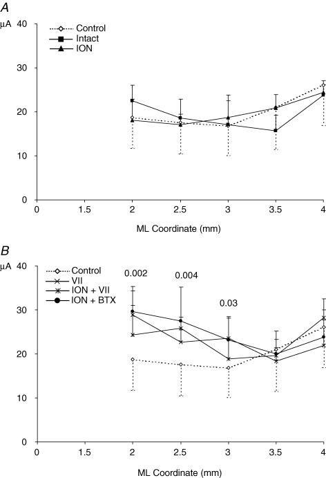
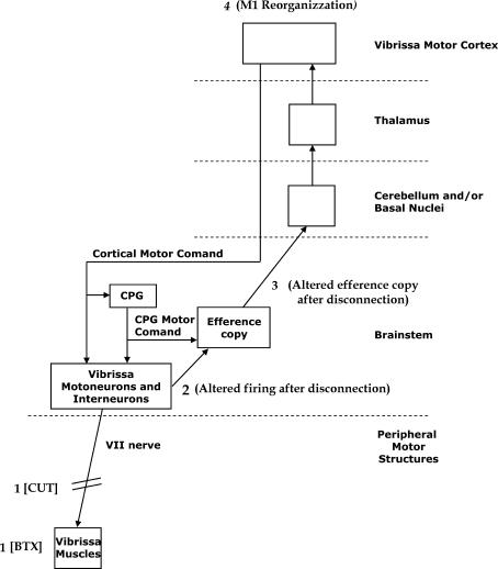
It has been proposed that abnormal vibrissae input to the motor cortex (M1) mediates short-term cortical reorganization after facial nerve lesion. To test this hypothesis, we cut first the infraorbital nerve (ION cut) and then the facial nerve (VII cut) in order to evaluate M1 reorganization without any aberrant, facial-nerve-lesion-induced sensory feedback. In each animal, M1 output was assessed in both hemispheres by mapping movements induced by intracortical microstimulation. M1 output was compared in different types of peripheral manipulations: (i) contralateral intact vibrissal pad (intact hemispheres), (ii) contralateral VII cut (VII hemispheres), (iii) contralateral ION cut (ION hemispheres), (iv) contralateral VII cut after contralateral ION cut (ION + VII hemispheres), (v) contralateral pad botulinum-toxin-injected after ION cut (ION + BTX hemispheres). Right and left hemispheres in untouched animals were the reference for normal M1 map (control hemispheres). Findings demonstrated that: (1) in ION hemispheres, the mean size of the vibrissae representation was not significantly different from those in intact and control hemispheres; (2) reorganization of the vibrissae movement representation clearly emerged only in hemispheres where the contralateral vibrissae pad had undergone motor output disconnection (VII cut hemispheres); (3) the persistent loss of vibrissae input did not change the M1 reorganization pattern during the first 48 h after motor paralysis (ION + VII cut and ION + BTX hemispheres). Thus, after motor paralysis, vibrissa input does not provide the gating signal necessary to trigger M1 reorganization.


Figures

Figure 1
Figure 1. Diagrams illustrating the experimental design and nomenclature
Animals were divided into three experimental groups (animal group) and a control group (untouched animals, not shown). Peripheral manipulation: VII cut, facial nerve lesion at the exit from stilomastoid foramen; ION cut, infraorbital nerve lesion at the exit from infraorbital fissure. BTXØ, single injection of botulinum toxin at a single site in the vibrissa pad. Animal groups: VII, unilateral facial nerve lesion, 5 rats; ION + VII, bilateral infraorbital nerve lesion, followed by unilateral facial nerve lesion after 2 weeks, 5 rats; ION + BTX, bilateral infraorbital nerve lesion, followed by unilateral BTX injection after 2 weeks, 5 rats. Cortical hemispheres: intact, hemisphere contralateral to the intact vibrissa pad; VII, hemisphere contralateral to the deefferented vibrissa pad; ION, hemisphere contralateral to the deafferented vibrissa pad; ION + VII, hemisphere contralateral to the deafferented and deefferented vibrissa pad; ION + BTX hemisphere, hemisphere contralateral to the deafferented and BTX-injected pad.
Figure 2
Figure 2. Cross-section of the infraorbital nerve in a control, and proximal to the acrylic stopper 2 weeks postinjury
Light photomicrograph (×200 magnification, osmium tetroxide and toluidine blue stain). Note evidence of Wallerian degeneration and/or perineural thickening in all ION fascicles (insert, ×1000 magnification).
Figure 3
Figure 3. Surface view map and mediolateral distribution of movements in a control group
A, example of representation of movements evoked at threshold current levels in the right and left hemispheres in control rats. The microelectrode was sequentially introduced to a depth of 1500 μm. Interpenetration distances were 500 μm. In these M1 mapping schemes, frontal poles are at the bottom. 0 corresponds to the bregma; numbers indicate rostral or caudal distance from the bregma or lateral distance from the midline. Movement evoked at one point is indicated by the following symbols: large filled square, contralateral vibrissae; open square, ipsilateral vibrissae; open diamond, forelimb; open cross, hindlimb; filled triangle, neck-upper trunk; filled star, jaw–tongue; filled circle, eye; small filled square, sites unresponsive at 60 μA; no symbol (within or at the border of the maps), penetration not performed due to presence of a large vessel. B, the mediolateral frequency distribution of penetrations eliciting vibrissa (filled square) and forelimb (open diamonds) movements in the right (continuous line) and left hemispheres (dashed line) of the control rats. For each hemisphere, penetrations were distributed into 0.5 mm bins extending from the midline (0 mm) to 4 mm lateral of the midline, irrespective of the anteroposterior coordinate. The number of penetrations falling into each bin was tailed and expressed as a percentage of the total number of penetrations for that movement (+ s.d.). Note that the vibrissa movement sites at 3 and 3.5 mm of mediolateral coordinate do not appear in the example of bilateral map shown above.
Figure 4
Figure 4. Surface view maps in a VII group
Examples of bilateral vibrissa maps from three rats (A, B and C) showing one hemisphere with the contralateral vibrissa pad with intact input–output condition (intact hemisphere) and the other with the contralateral vibrissa pad with facial-nerve-cut-induced motor paralysis (VII hemisphere). Intact hemispheres showed a large contralateral vibrissa movement representation. In the VII hemisphere there was evidence of persistent ipsilateral vibrissae and neck movements in the medial part of the former vibrissa representation. Moreover, it is worth noting that the forelimb expanded medially inside the former vibrissa representation.
Figure 5
Figure 5. Surface view maps in a ION + VII group.
Examples of bilateral vibrissa maps from three rats (A, B and C) showing one hemisphere with the contralateral vibrissae pad ION-cut-deafferented (ION hemisphere) and the other with contralateral VII-cut-induced paralysis after vibrissae pad ION-cut-deafferented (ION + VII hemisphere). ION hemispheres evidenced a large contralateral vibrissa movement representation similar to that found in the intact hemispheres in the Fig. 3. Also note that the pattern of represented movements in the ION + VII hemispheres were qualitatively similar to those in the VII hemispheres shown in Fig. 4.
Figure 6
Figure 6. Surface view maps in a ION + BTX group
Examples of bilateral vibrissa maps from three rats (A, B and C) showing one hemisphere with the contralateral vibrissae pad ION-cut-deafferented (ION hemisphere) and the other with the BTX-induced paralysis after ION cut in the contralatera vibrissa pad (ION + BTX hemisphere). The pattern of represented movement in the ION hemispheres showed a large contralateral vibrissa movement representation similar to that found in the ION hemispheres in the Fig. 5 and in the intact hemispheres in the Fig. 4. Also note that, in the ION + BTX hemispheres, the pattern of represented movements was qualitatively similar to that found in the VII hemispheres in the Fig. 4 and in the ION + VII hemispheres in the Fig. 5.
Figure 7
Figure 7. Percentage of unresponsive and effective sites devoted to various movements in each of the group of hemispheres studied
Across-group comparisons were made of unresponsive and effective sites devoted to various movements represented in M1 (up to 2.5 mm from the midline) for all hemispheres: control, intact, ION, VII, ION + VII, ION + BTX. Contralateral vibrissa movement is absent in deeferented groups and ipsilateral vibrissa movement is absent in the intact and ION hemispheres. In comparison with control and intact hemispheres, the ION hemisphere showed no significant difference in the percentage of unresponsive sites (A) or in the percentage of movement sites (B–F). In contrast, in comparison with control and intact hemispheres, the percentage of unresponsive sites (A), ipsilateral vibrissae (C), neck (D) and forelimb sites (E) was significantly increased in the VII, ION + VII and ION + BTX hemispheres. Comparison between the VII, ION + VII and ION + BTX hemispheres showed no significant difference in the percentage of unresponsive sites (A) or in the percentage of movement sites (B–F). Data are means + s.d. *Statistically significant differences between control versus ION, VII, ION + VII and ION + BTX hemispheres using the post hoc Scheffé test (S20.05;4,30 > 10.76; P < 0.05).The percentage of hindlimb movement sites is not reported because this movement was not extensively explored.
Figure 8
Figure 8. Distribution of forelimb movement sites in each of the group of hemispheres studied
Comparison of mediolateral frequency distribution of penetration eliciting forelimb movement between the control and the intact and ION hemispheres (A) and between the control and the VII, ION + VII and ION + BTX hemispheres (B). The mediolateral position of the forelimb sites was distributed in 0.5 mm bins extending from the midline to 4 mm lateral, irrespective of the anteroposterior coordinate. The number of sites at each bin was expressed as a percentage of the total forelimb sites. Note that, in the ION hemispheres, the percentage of 2 and 2.5 mm mediolateral coordinate sites overlap those of the control (A, P > 0.05, χ2 test); in contrast, at the same sites, the percentage was significantly increased in the VII, ION + VII, and ION + BTX hemispheres (B, P < 0.05, χ2 test). There is no difference in the frequency distribution between VII, ION + VII and ION + BTX hemispheres (P > 0.05, χ2 test). *Statistically significant differences between control versus VII, ION + VII and ION + BTX hemispheres using the χ2 test.
Figure 9
Figure 9. Threshold current level for eliciting forelimb movement in each of the group of hemispheres studied
In each group, the filled bar shows the threshold of forelimb sites situated 3.5–4 mm lateral to the midline (P = 0.94, ANOVA) and the open bar shows the threshold of forelimb sites 2–3 mm lateral (P < 0.0001, ANOVA). After vibrissae motor disconnection (VII, ION + VII and ION + BTX hemispheres) the threshold value increases only in the left bar containing the threshold current required to elicit the novel forelimb movement from the former vibrissae representation. There is no difference in the left bar value between control, intact and ION hemispheres (P > 0.05 Scheffé test) and between the VII, ION + VII and ION + BTX hemispheres (P > 0.05 Scheffé test). This evidence shows that the vibrissae input did not affect site excitability in the reorganizing portion motor cortex where novel forelimb movement emerges. *P < 0.05, Scheffé test: control, intact, ION versus VII, ION + BTX, ION + VII.
Figure 10
Figure 10. Distribution of thresholds for forelimb movement in each of the group of hemispheres studied
Mediolateral distribution of thresholds for forelimb movements: comparison of control versus intact and ION hemispheres (A) and of control versus VII, ION + VII and ION + BTX hemispheres (B). Thresholds were grouped according to their distance from the midline irrespective of the anteroposterior coordinate. The numbers above the graph lines (B) indicate the statistical probability derived by group comparison (ANOVA). This graph shows: (i) in the ION hemispheres the threshold values overlap with the values for the control and intact hemispheres throughout the mediolateral coordinate; (ii) in all three deefferented hemispheres (B) at 3, 2.5 and 2 mm laterally, the threshold value is higher than in control hemispheres and increases progressively; threshold values in the other more lateral sites overlap with those of the control hemispheres. In each mediolateral coordinate, the mean threshold value is reported only when the mean value was obtained in 5 animals for each group.
Figure 11
Figure 11. The proposed mechanisms of short-term reorganization of input-deprived motor vibrissa representation following motor disconnection
The facial nucleus motor neurons and interneurons receive cortical motor commands and central pattern generator (CPG) motor commands. A copy of the motor command, that is, an efference copy, ascends from the brainstem to the vibrissa motor cortex (further explanations in the Discussion). The vibrissa motor disconnection by VII cut or BTX injection (1) might induce changes in the firing of facial nucleus motor neurons and interneurons (2) that might change the efference copy ascending to the motor cortex (3) that might trigger M1 reorganization (4).

Similar articles

Cited by

References

    1. Ahrens KF, Kleinfeld D. Current flow in vibrissa motor cortex can phase-lock with exploratory rhythmic whisking in rat. J Neurophysiol. 2004;92:1700–1707. - PubMed
    1. Angaut P, Cicirata F. Anatomo-functional organization of the neocerebellar control pathways on the cerebral motor cortex. Rev Neurol. 1994;150:39–45. - PubMed
    1. Armstrong-James M, George MJ. Bilateral receptive fields of cells in rat Sm1 cortex. Exp Brain Res. 1988;70:155–165. - PubMed
    1. Asanuma H, Stoney SJ, Abzung C. Relationship between afferent input and motor outflow in cat motorsensory cortex. J Neurophysiol. 1968;31:670–681. - PubMed
    1. Berg WR, Kleinfeld D. Vibrissae movement elicited by rhythmic electrical microstimulation to motor cortex in the aroused rat mimics exploratory whisking. J Neurophysiol. 2003;90:2950–2963. - PubMed

LinkOut - more resources