. 2006 May 23;103(21):8060-5.
doi: 10.1073/pnas.0602606103. Epub 2006 May 11.

Toward the structural genomics of complexes: crystal structure of a PE/PPE protein complex from Mycobacterium tuberculosis

Affiliations

Toward the structural genomics of complexes: crystal structure of a PE/PPE protein complex from Mycobacterium tuberculosis

Michael Strong et al. Proc Natl Acad Sci U S A. .

Abstract

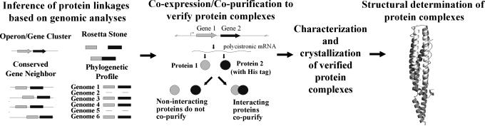
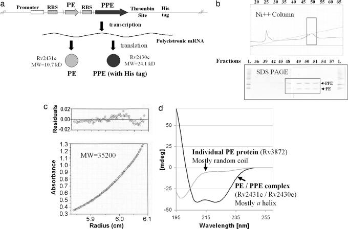
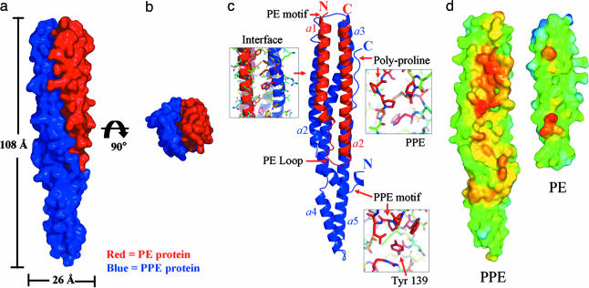
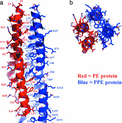
The developing science called structural genomics has focused to date mainly on high-throughput expression of individual proteins, followed by their purification and structure determination. In contrast, the term structural biology is used to denote the determination of structures, often complexes of several macromolecules, that illuminate aspects of biological function. Here we bridge structural genomics to structural biology with a procedure for determining protein complexes of previously unknown function from any organism with a sequenced genome. From computational genomic analysis, we identify functionally linked proteins and verify their interaction in vitro by coexpression/copurification. We illustrate this procedure by the structural determination of a previously unknown complex between a PE and PPE protein from the Mycobacterium tuberculosis genome, members of protein families that constitute approximately 10% of the coding capacity of this genome. The predicted complex was readily expressed, purified, and crystallized, although we had previously failed in expressing individual PE and PPE proteins on their own. The reason for the failure is clear from the structure, which shows that the PE and PPE proteins mate along an extended apolar interface to form a four-alpha-helical bundle, where two of the alpha-helices are contributed by the PE protein and two by the PPE protein. Our entire procedure for the identification, characterization, and structural determination of protein complexes can be scaled to a genome-wide level.


Conflict of interest statement

Conflict of interest statement: No conflicts declared.

Figures

Fig. 1.
Fig. 1.
Combined computational and biochemical procedure for the identification, characterization, and structural determination of protein complexes. Four computational methods are used to identify functionally linked proteins based on genomic analyses. Putative interacting proteins are cloned into a coexpression vector, where one protein is tagged with a His affinity tag and the others are not tagged. Genes are coexpressed, and interacting proteins are identified by affinity chromatography. If the proteins interact to form a long-lived protein complex, then the nontagged protein(s) copurifies with the tagged protein. Newly identified protein complexes are characterized and crystallized. We demonstrate this strategy by identifying, characterizing, and determining the crystal structure of a M.tb. PE/PPE protein complex.
Fig. 2.
Fig. 2.
Validation and characterization of the Rv2431c/Rv2430c PE/PPE protein complex. (a) The PE and PPE genes, Rv2431c and Rv2430c, were cloned and ligated into a coexpression vector. In this system the PPE protein is tagged with a His tag, whereas the PE protein is not tagged. Long-lived protein–protein interactions are identified by the coelution and copurification of the untagged PE protein with the tagged PPE protein. (b) Identification of interacting PE and PPE proteins. The soluble supernatant from the coexpressed PE and PPE experiments was bound to and eluted from a nickel affinity column. The untagged PE protein coelutes with the tagged PPE protein, suggesting a physical association of the two proteins. (c) Sedimentation equilibrium experiments suggested that the 10.7-kDa PE and 24.1-kDa His-tagged PPE proteins form a 1:1 heterodimer. (d) CD experiments show that the Rv2431c/Rv2430c PE/PPE protein complex is folded and mostly α-helical. This result is in contrast to the individually expressed PE protein, Rv3872, which is soluble but unfolded.
Fig. 3.
Fig. 3.
Crystal structure of the M.tb. PE/PPE protein complex. (a) Surface representation of the PE/PPE protein complex. The PE protein Rv2431c is shown in red, and the PPE protein Rv2430c is in blue. (b) The PE/PPE protein complex viewed down its longitudinal axis. (c) Ribbon diagram of the PE/PPE protein complex. The complex is composed of seven α-helices. Two α-helices of the PE protein interact with two helices of the PPE protein to form a four-helix bundle. Regions of high sequence conservation are indicated by arrows and discussed in the text. (d) Interface hydrophobicity of the PPE and PE proteins. The hydrophobicity of the interaction interface between the PPE and PE protein is color-coded: the most apolar regions are indicated in red, orange, and yellow, and the most polar regions are indicated in blue. Notice the extensive apolar regions that are shielded from solvent as the complex forms.
Fig. 4.
Fig. 4.
Interaction interface of the PE/PPE protein complex. (a) Two helices of the PE protein, α1 and α2, interact with two helices of the PPE protein, α2 and α3, to form a four-helix bundle. The four-helix-bundle is largely stabilized by hydrophobic interactions among apolar side chains, as seen in the core of the complex. In contrast, the outer surface is coated with polar residues. (b) The PE/PPE complex as viewed down the longitudinal axis.

References

    1. Uetz P., Giot L., Cagney G., Mansfield T. A., Judson R. S., Knight J. R., Lockshon D., Narayan V., Srinivasan M., Pochart P., et al. Nature. 2000;403:623–627. - PubMed
    1. Ito T., Chiba T., Ozawa R., Yoshida M., Hattori M., Sakaki Y. Proc. Natl. Acad. Sci. USA. 2001;98:4569–4574. - PMC - PubMed
    1. Gavin A. C., Bosche M., Krause R., Grandi P., Marzioch M., Bauer A., Schultz J., Rick J. M., Michon A. M., Cruciat C. M., et al. Nature. 2002;415:141–147. - PubMed
    1. Ho Y., Gruhler A., Heilbut A., Bader G. D., Moore L., Adams S. L., Millar A., Taylor P., Bennett K., Boutilier K., et al. Nature. 2002;415:180–183. - PubMed
    1. Rual J. F., Venkatesan K., Hao T., Hirozane-Kishikawa T., Dricot A., Li N., Berriz G. F., Gibbons F. D., Dreze M., Ayivi-Guedehoussou N., et al. Nature. 2005;437:1173–1178. - PubMed

Publication types