. 2006 May 23;103(21):8090-5.
doi: 10.1073/pnas.0601083103. Epub 2006 May 11.

Oogenesis requires germ cell-specific transcriptional regulators Sohlh1 and Lhx8

Affiliations

Oogenesis requires germ cell-specific transcriptional regulators Sohlh1 and Lhx8

Stephanie A Pangas et al. Proc Natl Acad Sci U S A. .

Abstract

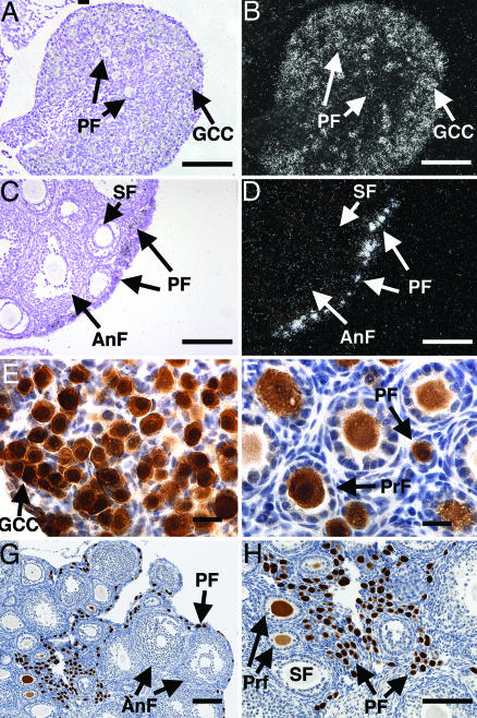
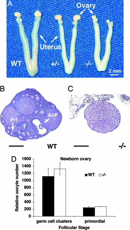
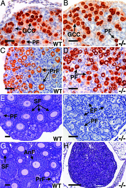
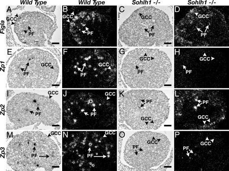
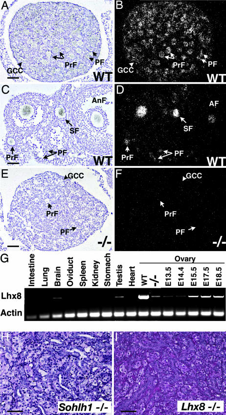
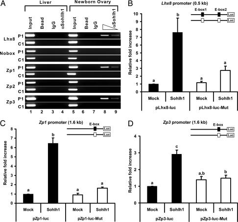
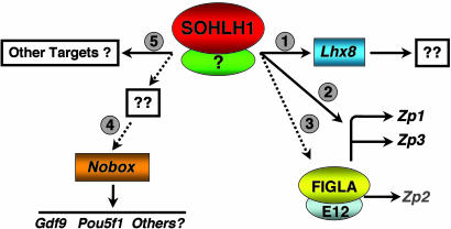
Mammalian oogenesis requires oocyte-specific transcriptional regulators. The full complement of oocyte-specific transcription factors is unknown. Here, we describe the finding that Sohlh1, a spermatogenesis and oogenesis basic helix-loop-helix transcription factor in females, is preferentially expressed in oocytes and required for oogenesis. Sohlh1 disruption perturbs follicular formation in part by causing down-regulation of two genes that are known to disrupt folliculogenesis: newborn ovary homeobox gene (Nobox) and factor in the germ-line alpha (Figla). In addition, we show that Lhx8 is downstream of Sohlh1 and critical in fertility. Thus, Sohlh1 and Lhx8 are two germ cell-specific, critical regulators of oogenesis.


Conflict of interest statement

Conflict of interest statement: No conflicts declared.

Figures

Fig. 1.
Fig. 1.
Sohlh1 mRNA and protein expression. (AD) In situ hybridization with Sohlh1 riboprobe on newborn (A and B) and 6-week-old (C and D) ovarian tissue. Bright-field (A and C) and dark-field (B and D) views are shown. Arrows throughout show locations of germ cell cysts (GCC), primordial follicles (PF), primary follicles (PrF), secondary follicles (SF), and antral follicles (AnF). Sohlh1 transcripts localize mainly to germ cell cysts and oocytes in primordial follicles. (EH) Rabbit antibodies against SOHLH1 were used to perform immunohistochemistry on newborn (E), 9-day-old (F), and 6-week-old (G and H) ovaries. Immunoreactivity to the SOHLH1 protein stained brown, and cell nuclei were counterstained blue with hematoxylin. (Scale bars: AD and G and H, 100 μm; E and F, 20 μm.)
Fig. 2.
Fig. 2.
Sohlh1 adult knockout anatomy, histology, and histomorphometric analysis. (A) Gross reproductive tracts dissected from WT, heterozygous (+/−), and homozygous (−/−) Sohlh1 mice. Note markedly smaller ovaries in Sohlh1−/− mice. (B) WT ovary with advanced antral follicle (AnF) as well as primary follicles (Prf). (C) Sohlh−/− ovary (−/−) lacks germ cells. (D) Five pairs of newborn ovaries from WT and Sohlh1 knockout (−/−) mice were sectioned, and oocytes within the germ cell clusters and primordial follicles were counted. No significant differences were observed between the WT and knockout ovaries. Data are represented as mean values, with error bars representing the SEM. Fisher's exact t test was used to calculate P values. (Scale bars: 400 μm.)
Fig. 3.
Fig. 3.
Sohlh1 knockout histology and immunohistochemistry; WT and knockout (−/−) data are shown. (A and B) Newborn ovaries stained with antibodies against GCNA1 show no difference in primordial follicles (PF) or germ cell cysts (GCC) between WT (A) and knockout (B). (C and D) Three-day ovaries stained anti-MSY2. Primary follicles (PrF) are seen in WT (C) but not knockout (D) ovaries. (E and F) Periodic acid/Schiff reagent (PAS) staining of 7-day WT (E) and knockout (F) ovaries show fewer follicle types and empty follicles (EF) in the knockout (F). (G and H) PAS staining of 3-week ovaries shows no remaining oocytes in the knockout (H) but all stages of development in the WT (G). (Scale bars: AG, 50 μm; H, 400 μm.)
Fig. 4.
Fig. 4.
Expression of Figla, Zp1, Zp2, and Zp3 in WT and Sohlh1−/− ovaries. Bright-field (A, C, E, G, I, K, M, and O) and their corresponding dark-field (B, D, F, H, J, L, N, and P) images of in situ hybridization from WT (A, B, E, F, I, J, M, and N) and Sohlh1−/− (C, D, G, H, K, L, O, and P) newborn ovaries. In newborn WT ovaries, Figla (A and B), Zp1 (E and F), Zp2 (I and J), and Zp3 (M and N) are expressed in germ cell cysts (GCC; arrowheads) and primordial follicles (PF; arrows). Expression of Figla (C and D) and Zp2 (K and L) are detectable in Sohlh1−/− ovaries by in situ hybridization. Zp1 (G and H) and Zp3 (O and P) are not detectable by in situ hybridization in Sohlh1−/− ovaries. Magnification is the same in AP. (Scale bars: 40 μm.)
Fig. 5.
Fig. 5.
Lhx8 expression and ovarian phenotype. (AF) Bright-field (A, C, and E) and dark-field (B, D, and F) views of in situ hybridization are shown with Lhx8 riboprobe to WT newborn (A and B) and 6-week-old (C and D) ovaries. (E and F) The Lhx8 riboprobe showed no significant hybridization to Sohlh1−/− ovaries. (G) Oligonucleotides corresponding to Lhx8 amplified RNA in WT testes and newborn ovaries (WT) but showed a dramatic decrease in Sohlh1−/− ovaries (−/−). Total RNA from embryonic ovaries (E13.5, E14.5, E15.5, E17.5, and E18.5) was isolated and also amplified with Lhx8-specific primers. (H and I) Periodic acid/Schiff reagent staining of 12-week old (adult) ovaries from Sohlh1−/− (H) and Lhx8−/− (I) shows a lack of germ cells in both mutants. GCC, germ cell cyst; PF, primordial follicle; PrF, primary follicle; SF, secondary follicle; AnF, antral follicle. (Scale bars: 40 μm.)
Fig. 6.
Fig. 6.
SOHLH1 binding and transactivation of the Lhx8, Zp1, and Zp3 promoters. (A) ChIP assays with anti-SOHLH1 antibodies on newborn ovary and liver extracts. Anti-SOHLH1 antibodies precipitate genomic DNA containing conserved E boxes from Lhx8, Zp1, and Zp3 promoter regions (P1) but not control genomic regions (C1). PCR amplifications of the E boxes are described in the legend of Fig. 12 and Table 2. “Input” is PCR product from chromatin pellets before immunoprecipitation. Samples incubated with anti-SOHLH1 antibody (αSohlh1) and the control sample without antibody (Bead) or IgG were used as templates for PCR. (BD) Transient transfection analyses of Lhx8 (B), Zp1 (C), and Zp3 (D) promoter regions with SOHLH1. Reporter constructs containing the WT (filled box, pLhx8-luc, pZp1-luc, and pZp3-luc) or mutant E boxes (open box, pLhx8-luc-Mut, pZp1-luc-Mut, and pZp3-luc-Mut) were cotransfected with vector expressing SOHLH1 or the empty vector (Mock). Lhx8 putative promoter contains two E boxes, and both were mutated. Conserved E boxes and mutated sequences are shown in Fig. 12 and Table 1. The mean fold increase in luciferase activity (± SEM) of triplicate experiments relative to the empty vector is shown. Statistical significance was determined by one-way ANOVA followed by the Tukey–Kramer honestly significant difference test for multiple comparisons. Bars marked with difference letters (a, b, and c) indicate statistical significance (P < 0.001).
Fig. 7.
Fig. 7.
Hypothetical model for SOHLH1 role in early folliculogenesis. SOHLH1 regulates a number of genes that are required for early oocyte development. The SOHLH1 interacting partner is unknown. (Step 1) The homeobox gene Lhx8 is likely a direct transcriptional target of SOHLH1, but genes downstream of LHX8 in the oocyte are unknown. (Step 2) Zp1 and Zp3 are likely directly regulated by SOHLH1, possibly in conjunction with FIGLA. (Step 3) Figla is partially down-regulated in Sohlh1 null oocytes, but Zp2, which is controlled by the FIGLA/E12 complex, is not significantly changed. (Step 4) The oocyte-specific homeobox gene, Nobox, is downstream of SOHLH1. Loss of Nobox expression in Sohlh1 null oocytes also results in loss of genes downstream of the NOBOX pathway, such as Gdf9 and Pou5f1. (Step 5) It is likely that SOHLH1 has additional target genes. Dotted lines indicate unknown pathways. Solid lines indicate direct transcriptional regulation.

References

    1. Pepling M. E., Spradling A. C. Dev. Biol. 2001;234:339–351. - PubMed
    1. Wylie C. Cell. 1999;96:165–174. - PubMed
    1. Rajkovic A., Pangas S. A., Ballow D., Suzumori N., Matzuk M. M. Science. 2004;305:1157–1159. - PubMed
    1. Dong J., Albertini D. F., Nishimori K., Kumar T. R., Lu N., Matzuk M. M. Nature. 1996;383:531–535. - PubMed
    1. Dean J. J. Reprod. Immunol. 2002;53:171–180. - PubMed

Publication types

Substances