

Selenoprotein deficiency accelerates prostate carcinogenesis in a transgenic model
- PMID: 16690748
- PMCID: PMC1472449
- DOI: 10.1073/pnas.0508218103
Selenoprotein deficiency accelerates prostate carcinogenesis in a transgenic model
Abstract
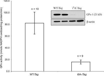
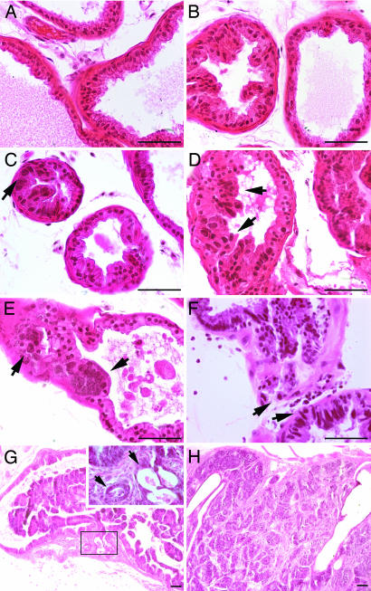
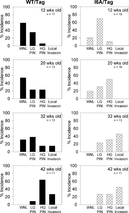
Considerable animal and human data have indicated that selenium is effective in reducing the incidence of several different types of cancer, including that of the prostate. However, the mechanism by which selenium inhibits carcinogenesis remains unknown. One possibility is that dietary selenium influences the levels of selenium-containing proteins, or selenoproteins. Selenoproteins contain selenium in the form of selenocysteine and perform a variety of cellular functions, including antioxidant defense. To determine whether the levels of selenoproteins can influence carcinogenesis independent of selenium intake, a unique mouse model was developed by breeding two transgenic animals: mice with reduced selenoprotein levels because of the expression of an altered selenocysteine-tRNA (i6A-) and mice that develop prostate cancer because of the targeted expression of the SV40 large T and small t oncogenes to that organ [C3(1)/Tag]. The resulting bigenic animals (i6A-/Tag) and control WT/Tag mice were assessed for the presence, degree, and progression of prostatic epithelial hyperplasia and nuclear atypia. The selenoprotein-deficient mice exhibited accelerated development of lesions associated with prostate cancer progression, implicating selenoproteins in cancer risk and development and raising the possibility that selenium prevents cancer by modulating the levels of these selenoproteins.
Conflict of interest statement
Conflict of interest statement: No conflicts declared.
Figures
References
-
- Clark L. C., Combs G. F., Turnbull B. W., Slate E. H., Chalker E. H., Chow J., Davis L. S., Glover R. A., Graham G. F., Gross E. G., et al. J. Am. Med. Assoc. 1996;276:1957–1963. - PubMed
-
- Yoshizawa K., Willett W. C., Morris S. J., Stampfer M. J., Speigelman D., Rimm E. B., Giovannucci E. J. Natl. Cancer Inst. 1998;90:1219–1224. - PubMed
-
- Li H., Stampfer M. J., Giovannucci E. L., Morris J. S., Willett W. C., Gaziona M., Ma J. J. Natl. Cancer Inst. 2004;96:696–703. - PubMed
-
- van den Brandt P. A., Zeegers M. P. A., Bode P., Goldbohm R. A. Cancer Epidemiol. Biomarkers Prevention. 2003;12:866–871. - PubMed
-
- Lippman S. M., Goodman P. J., Klein E. A., Parnes H. L., Thompson I. M., Jr., Kristal A. R., Santella R. M., Probstfield J. L., Moinpour C. M., Albanes D., et al. J. Natl. Cancer Inst. 2005;97:94–102. - PubMed
Publication types
MeSH terms
Substances
Grants and funding
LinkOut - more resources
Full Text Sources
Other Literature Sources
Medical
Molecular Biology Databases
Miscellaneous
