

doi: 10.1016/j.vibspec.2005.02.015.
Infrared spectroscopic characterization of mineralized tissues
- PMID: 16691288
- PMCID: PMC1459415
- DOI: 10.1016/j.vibspec.2005.02.015
Item in Clipboard
Infrared spectroscopic characterization of mineralized tissues
Vib Spectrosc.
.
Abstract
Vibrational spectroscopy (Infrared and Raman), and in particular micro-spectroscopy and micro-spectroscopic imaging has been used to characterize developmental changes in bone and other mineralized tissues, to monitor these changes in cell cultures, and to detect disease and drug-induced modifications. Examples of the use of infrared micro-spectroscopy and micro-spectroscopic imaging are discussed in this review.
Figures
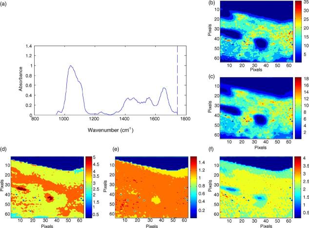
Hyperspectral images of bone mineral properties: in normal human cortical bone (a) typical spectrum from a single image pixel, (b) image of the mineral distribution in the biopsy, (c) image of the matrix distribution in the biopsy, (d) image of carbonate distribution, (e) image of mineral:matrix ratio, (f) image of crystallinity and (g) image of collagen cross link ratio. Note: all images are corrected for the presence of the embedding media, PMMA.
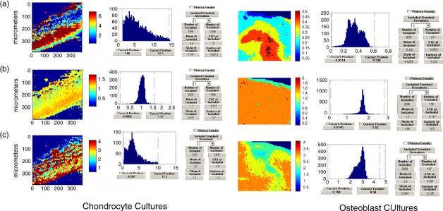
Images of mineralizing chondrocyte (left) and osteoblast (right) cultures: (a) mineral:matrix ratio, (b) crystallinity and (c) collagen cross-link ratio.
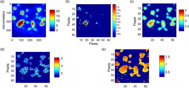
Image of a pathologic deposit removed from a patient with juvenile dermatomyositis: (a) mineral distribution, (b) lipid distribution, (c) mineral:matrix ratio (d) collagen cross-link ratio and (e) crystallinity distribution.

Pixel historgrams for the average distribution of mineral:matrix, crystallinity and collagen cross link ratio in normal and osteoporotic biopsies.
References
-
- Grant GA, Wener MH, Yaziji H, Futran N, Bronner MP, Mandel N, Mayberg MR. Destructive tophaceous calcium hydroxyapatite tumor of the infratemporal fossa. Case report and review of the literature. J Neurosurg. 1999;90(1):148–152. - PubMed
-
- Derfus BA, Rachow JW, Mandel NS, Boskey AL, Buday M, Kushnaryov VM, Ryan LM. Articular cartilage vesicles generate calcium pyrophosphate dihydrate-like crystals in vitro. Arthritis Rheum. 1992;35(2):231–240. - PubMed
-
- Feinberg J, Boachie-Adjei O, Bullough PG, Boskey AL. The distribution of calcific deposits in intervertebral discs of the lumbosacral spine. Clin. Orthop. 1990;(254):303–310. - PubMed
-
- Arlet J, Legros R, Savio JL, Bonel G. Crystallographic identification of a calcium deposit in calcified pericarditis associated with articular chondrocalcinosis. Bone. 1986;7(3):187–191. - PubMed
-
- Li C, Ebenstein D, Xu C, Chapman J, Sal oner D, Rapp J, Pruitt L. Biochemical characterization of atherosclerotic plaque constituents using FTIR spectroscopy and histology. J. Biomed. Mater. Res. 2003;64A(2):197–206. - PubMed
Grants and funding
LinkOut - more resources
Full Text Sources
Other Literature Sources