

Identification and quantification of a guanine-thymine intrastrand cross-link lesion induced by Cu(II)/H2O2/ascorbate
- PMID: 16696563
- PMCID: PMC2519820
- DOI: 10.1021/tx060025x
Identification and quantification of a guanine-thymine intrastrand cross-link lesion induced by Cu(II)/H2O2/ascorbate
Abstract
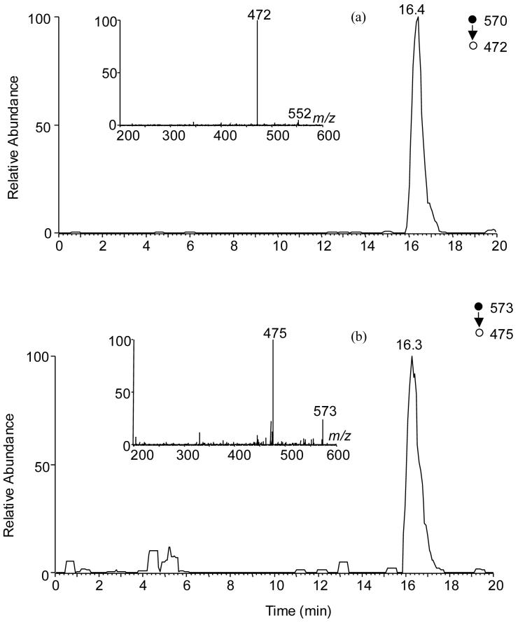
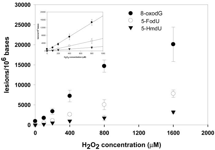
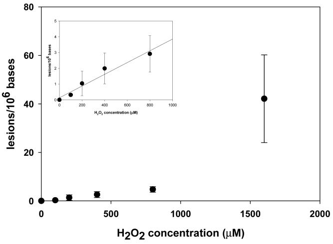
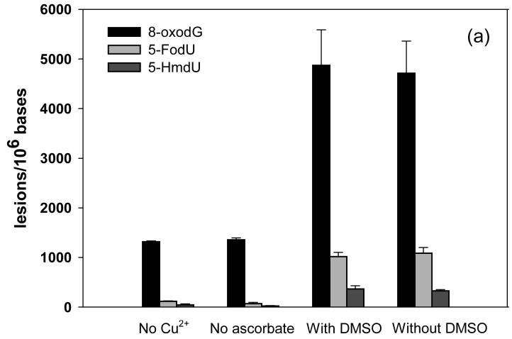
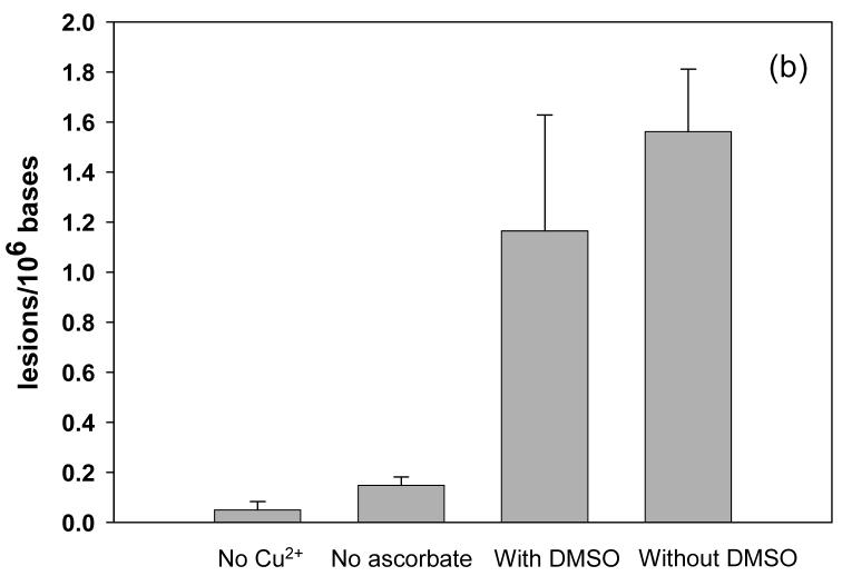
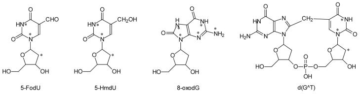
Reactive oxygen species (ROS) can be induced by both endogenous and exogenous processes, and they can damage biological molecules including nucleic acids. It was shown that X- or gamma-ray irradiation of aqueous solutions of DNA, during which *OH is one of the major ROS, can lead to the formation of intrastrand cross-link lesions where the neighboring nucleobases in the same DNA strand are covalently bonded. Previous 32P-postlabeling studies suggested that the intrastrand cross-link lesions may arise from Fenton reaction, which also induces the formation of *OH; the structures of the proposed intrastrand cross-link lesions, however, have not been determined. Here, we showed for the first time that the treatment of calf thymus DNA with Cu(II)/H2O2/ascorbate could lead to the formation of an intrastrand cross-link lesion, i.e., G wedge T, where the C8 of guanine is covalently bonded to the neighboring 3'-thymine through its methyl carbon. LC-MS/MS quantification results showed dose-responsive formation of G wedge T. In addition, the yield of the intrastrand cross-link was approximately 3 orders of magnitude lower than those of commonly observed single-base lesions, that is, 8-oxo-7,8-dihydro-2'-deoxyguanosine, 5-(hydroxymethyl)-2'-deoxyuridine, and 5-formyl-2'-deoxyuridine. The induction of intrastrand cross-link lesion in calf thymus DNA by Fenton reagents in vitro suggests that this type of lesion might be formed endogenously in mammalian cells.
Figures
References
-
- Lindahl T. DNA lesions generated in vivo by reactive oxygen species, their accumulation and repair. NATO ASI Ser., Ser. A. 1999;302:251–257.
-
- Finkel T, Holbrook NJ. Oxidants, oxidative stress and the biology of ageing. Nature. 2000;408:239–247. - PubMed
-
- Marnett LJ. Oxyradicals and DNA damage. Carcinogenesis. 2000;21:361–370. - PubMed
-
- Feig DI, Reid TM, Loeb LA. Reactive oxygen species in tumorigenesis. Cancer Res. 1994;54:1890s–1894s. - PubMed
Publication types
MeSH terms
Substances
Grants and funding
LinkOut - more resources
Full Text Sources
Other Literature Sources
Medical
