. 2006 May;19(5):645-54.
doi: 10.1021/tx0600088.

Cross-linking of the human DNA repair protein O6-alkylguanine DNA alkyltransferase to DNA in the presence of 1,2,3,4-diepoxybutane

Affiliations

Cross-linking of the human DNA repair protein O6-alkylguanine DNA alkyltransferase to DNA in the presence of 1,2,3,4-diepoxybutane

Rachel Loeber et al. Chem Res Toxicol. 2006 May.

Abstract

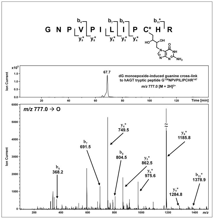
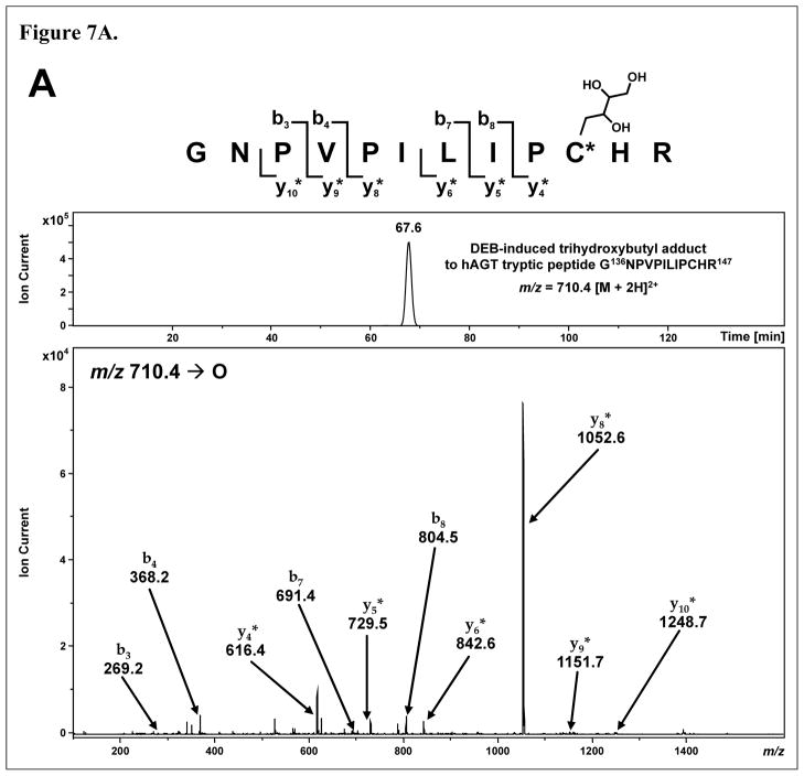
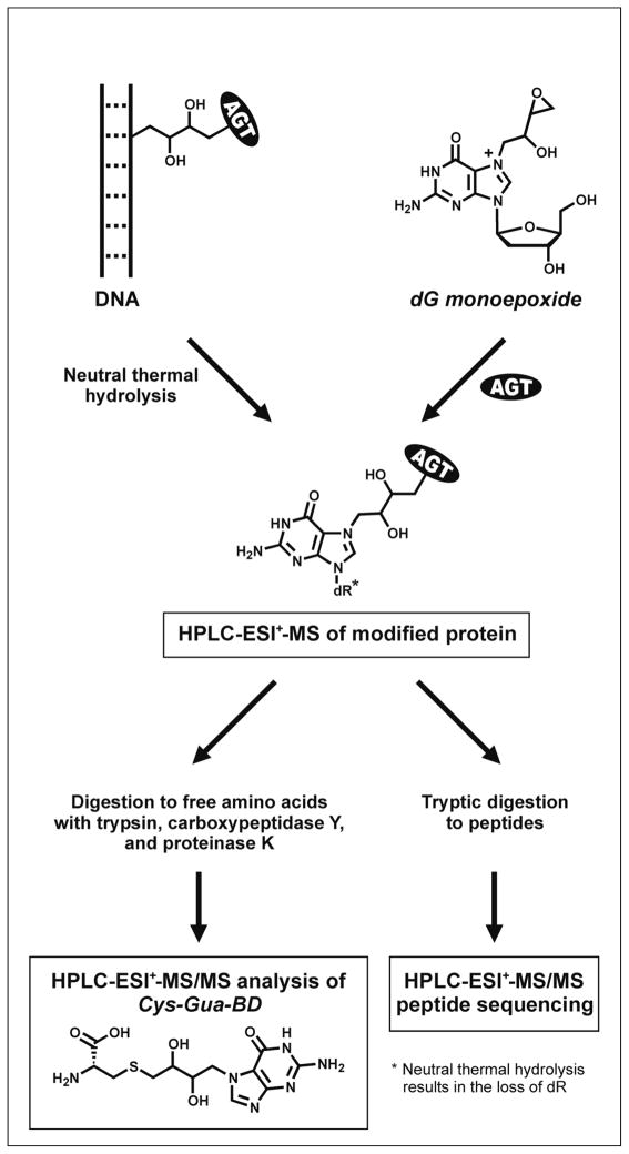
1,2,3,4-Diepoxybutane (DEB) is a key carcinogenic metabolite of the important industrial chemical 1,3-butadiene. DEB is a bifunctional alkylating agent capable of reacting with DNA and proteins. Initial DNA alkylation by DEB produces N7-(2'-hydroxy-3',4'-epoxybut-1'-yl)-guanine monoadducts, which can react with another nucleophilic site to form cross-linked adducts. A recent report revealed a strong correlation between cellular expression of the DNA repair protein O6-alkylguanine DNA alkyltransferase (AGT) and the cytotoxic and mutagenic activity of DEB, suggesting that DEB induces AGT-DNA cross-links (Valadez, J. G., et al. (2004) Activation of bis-electrophiles to mutagenic conjugates by human O6-alkylguanine-DNA alkyltransferase. Chem. Res. Toxicol. 17, 972-982). The purpose of our study was to analyze the formation and structures of DEB-induced AGT-DNA conjugates and to identify specific amino acid residues within the protein involved in cross-linking. DNA-protein cross-link formation was detected by SDS-PAGE when 32P-labeled double-stranded oligodeoxynucleotides were exposed to DEB in the presence of either wild-type hAGT or a C145A hAGT mutant. Capillary HPLC-electrospray ionization mass spectrometry (ESI-MS) analysis of hAGT that had been treated with N7-(2'-hydroxy-3',4'-epoxybut-1'-yl)-deoxyguanosine (dG monoepoxide) revealed the ability of the protein to form either one or two butanediol-dG cross-links, corresponding to mass shifts of +353 and +706 Da, respectively. HPLC-ESI+ -MS/MS sequencing of the tryptic peptides obtained from dG monoepoxide-treated protein indicated that the two cross-linking sites were the alkyl acceptor site, Cys145, and a neighboring active site residue, Cys150. The same two amino acid residues of hAGT became covalently cross-linked to DNA following DEB treatment. Modification of Cys145 was further confirmed by HPLC-ESI+ -MS/MS analysis of dG monoepoxide-treated synthetic peptide GNPVPILIPCHR which represents the active site tryptic fragment of hAGT (C = Cys145). The replacement of the catalytic cysteine residue with alanine in the C145A hAGT mutant abolished DEB-induced cross-linking at this site, while the formation of conjugates via neighboring Cys150 was retained. The exact chemical structure of the cross-linked lesion was established as 1-(S-cysteinyl)-4-(guan-7-yl)-2,3-butanediol by HPLC-ESI+ -MS/MS analysis of the amino acids resulting from the total digestion of modified proteins analyzed in parallel with an authentic standard. AGT-DNA cross-linking is a likely mechanism of DEB-mediated cytotoxicity in cells expressing this important repair protein.


Figures

Figure 1
Figure 1
12% SDS-PAGE analysis of 32P-end labeled DNA duplexes (5′-GGA GCT GGT GGC GTA GGC-3′, (+) strand) following incubation with DEB and hAGT (lanes 4–6) or C145A hAGT (lane 7). AGT-DNA cross-links are observed as slowly moving bands on the gel. Lanes 1–3 serve as negative controls.
Figure 2
Figure 2
HPLC-ESI+-MS analysis of dG monoepoxide-induced butanediol cross-links to hAGT. (A) HPLC-ESI+-MS of untreated hAGT. Top: Total ion chromatogram; Bottom: ESI+ mass spectrum of the 16.6 min protein peak; Inset: Deconvoluted mass spectrum of the 16.6 min peak (observed M = 21 880 Da, calculated M = 21 876 Da). (B) HPLC-ESI+-MS of hAGT following incubation with 50 molar equivalents dG monoepoxide. Top: Total ion chromatogram; Bottom: ESI+ mass spectrum of the 16.3 min protein peak; Inset: Deconvoluted mass spectrum of the 16.3 min peak: A = hAGT containing a single cross-link to dG (observed M = 22 230 Da, calculated M = 22 229 Da); B = double cross-link to dG (observed M = 22 586 Da, calculated M = 22 582 Da); C = unmodified hAGT (observed M = 21 877 Da, calculated M = 21 876 Da).
Figure 3
Figure 3
HPLC-ESI+-MS analysis of C145A hAGT protein following treatment with dG monoepoxide. (A) HPLC-ESI+-MS of untreated C145A hAGT. Top: Total ion chromatogram; Bottom: ESI+ mass spectrum of the 16.2 min protein peak; Inset: Deconvoluted mass spectrum of the 16.2 min peak (observed M = 23 015 Da, calculated M = 23 012 Da). (B) HPLC-ESI+-MS of C145A hAGT mutant following incubation dG monoepoxide. Top: Total ion chromatogram; Bottom: ESI+ mass spectrum of the 16.1 min protein peak; Inset: Deconvoluted mass spectrum of the 16.1 min peak: A = C145A hAGT containing a single cross-link to dG (observed M = 23 367 Da, calculated M = 23 365 Da); B = unmodified protein (observed M = 23 014 Da, calculated M = 23 012 Da).
Figure 4
Figure 4
HPLC-ESI+-MS/MS analysis of hAGT tryptic peptide G136NPVPILIPCHR147 containing a dG monoepoxide-induced butanediol cross-link between Cys145 and guanine. Top: Extracted ion chromatogram of m/z 777.0 [M + 2H]2+; Bottom: MS/MS spectrum. Modified fragment ions are indicated by “*”.
Figure 5
Figure 5
HPLC-ESI+-MS/MS analysis of hAGT tryptic peptide V148VCSSGGAVGNYSG GLAVK165 containing dG monoepoxide-induced butanediol cross-link between Cys150 and guanine. Top: Extracted ion chromatogram of m/z 953.6 [M + 2H]2+; Bottom: MS/MS spectrum. Modified fragment ions are indicated by “*”.
Figure 6
Figure 6
HPLC-ESI+-MS/MS analysis of hAGT tryptic peptides containing DEB-induced butanediol cross-links to guanine. (A) Top: Extracted ion chromatogram of hAGT tryptic peptide G136NPVPILIPCHR147 cross-linked to guanine (m/z 776.9 [M + 2H]2+); Bottom: MS/MS spectrum of tryptic peptide G136NPVPILIPCHR147 mapping the cross-link to Cys145. (B) Top: Extracted ion chromatogram of hAGT tryptic peptide V148VCSSGGAVGNYSGGLAVK165 cross-linked to guanine (m/z 952.9 [M + 2H]2+); Bottom: MS/MS spectrum of tryptic peptide V148VCSSGGAVGNYSGGLAVK165 mapping the cross-link to Cys150. Modified fragment ions are indicated by “*”.
Figure 6
Figure 6
HPLC-ESI+-MS/MS analysis of hAGT tryptic peptides containing DEB-induced butanediol cross-links to guanine. (A) Top: Extracted ion chromatogram of hAGT tryptic peptide G136NPVPILIPCHR147 cross-linked to guanine (m/z 776.9 [M + 2H]2+); Bottom: MS/MS spectrum of tryptic peptide G136NPVPILIPCHR147 mapping the cross-link to Cys145. (B) Top: Extracted ion chromatogram of hAGT tryptic peptide V148VCSSGGAVGNYSGGLAVK165 cross-linked to guanine (m/z 952.9 [M + 2H]2+); Bottom: MS/MS spectrum of tryptic peptide V148VCSSGGAVGNYSGGLAVK165 mapping the cross-link to Cys150. Modified fragment ions are indicated by “*”.
Figure 7
Figure 7
HPLC-ESI+-MS/MS analysis of hAGT tryptic peptides G136NPVPILIPCHR147 and V148VCSSGGAVGNYSGGLAVK165containing DEB-induced 2′,3′,4′-trihydroxybut-1′-yl adducts to active site cysteine residues Cys145 (A) and Cys150 (B). Top: Extracted ion chromatograms; Bottom: MS/MS spectra. Modified fragment ions are indicated by “*”.
Figure 7
Figure 7
HPLC-ESI+-MS/MS analysis of hAGT tryptic peptides G136NPVPILIPCHR147 and V148VCSSGGAVGNYSGGLAVK165containing DEB-induced 2′,3′,4′-trihydroxybut-1′-yl adducts to active site cysteine residues Cys145 (A) and Cys150 (B). Top: Extracted ion chromatograms; Bottom: MS/MS spectra. Modified fragment ions are indicated by “*”.
Figure 8
Figure 8
HPLC-ESI+-MS/MS analysis of 1-(S-cysteinyl)-4-(guan-7-yl)-2,3-butanediol (Cys-Gua-BD) in total digests of AGT protein treated with dG monoepoxide. (A) Extracted ion chromatogram of Cys-Gua-BD (m/z 359.7 [M + H]+) resulting from the total digestion of dG monoepoxide-treated hAGT; Inset: MS/MS fragmentation of Cys-Gua-BD. (B) Extracted ion chromatogram of Cys-Gua-BD (m/z 359.4 [M + H]+) resulting from the total digestion of dG monoepoxide-treated C145A hAGT; Inset: MS/MS fragmentation of Cys-Gua-BD.
Scheme 1
Scheme 1
DNA-AGT cross-linking by dihaloethanes and DEB.
Scheme 2
Scheme 2
Mass spectrometric analysis of DEB-induced AGT-DNA conjugates.

References

    1. Pegg AE. Repair of O6-alkylguanine by alkyltransferases. Mutat Res. 2000;462:83–100. - PubMed
    1. Daniels DS, Woo TT, Luu KX, Noll DM, Clarke ND, Pegg AE, Tainer JA. DNA binding and nucleotide flipping by the human DNA repair protein AGT. Nat Struct Mol Biol. 2004;11:714–720. - PubMed
    1. Rasimas JJ, Dalessio PA, Ropson IJ, Pegg AE, Fried MG. Active-site alkylation destabilizes human O6-alkylguanine DNA alkyltransferase. Protein Sci. 2004;13:301–305. - PMC - PubMed
    1. Daniels DS, Mol CD, Arvai AS, Kanugula S, Pegg AE, Tainer JA. Active and alkylated human AGT structures: a novel zinc site, inhibitor and extrahelical base binding. EMBO J. 2000;19:1719–1730. - PMC - PubMed
    1. Federwisch M, Hassiepen U, Bender K, Dewor M, Rajewsky MF, Wollmer A. Recombinant human O6-alkylguanine-DNA alkyltransferase (AGT), Cys145-alkylated AGT and Cys145 --> Met145 mutant AGT: comparison by isoelectric focusing, CD and time-resolved fluorescence spectroscopy. Biochem J. 1997;324 (Pt 1):321–328. - PMC - PubMed

Publication types