

Effect of porcine respiratory coronavirus infection on lipopolysaccharide recognition proteins and haptoglobin levels in the lungs
- PMID: 16697680
- PMCID: PMC7110855
- DOI: 10.1016/j.micinf.2006.01.009
Effect of porcine respiratory coronavirus infection on lipopolysaccharide recognition proteins and haptoglobin levels in the lungs
Abstract
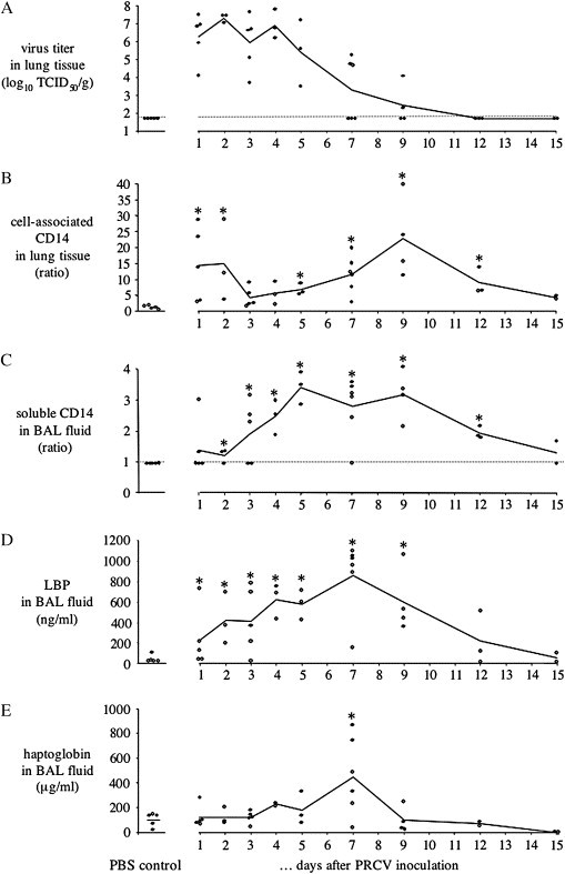
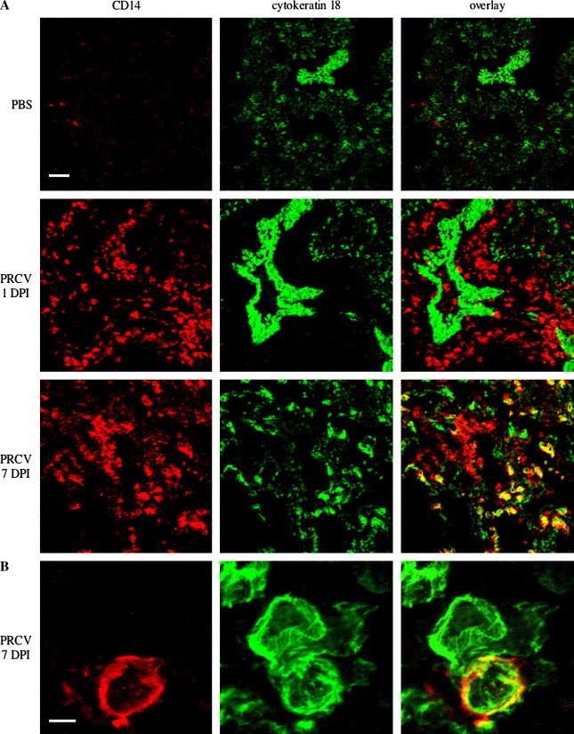
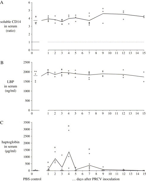
Porcine respiratory coronavirus (PRCV) potentiates respiratory disease and proinflammatory cytokine production in the lungs upon intratracheal inoculation with lipopolysaccharide (LPS) at 1 day of infection. This study aimed to quantify LPS-binding protein (LBP), CD14 and haptoglobin in the lungs throughout a PRCV infection. LBP and CD14 recognize LPS and enhance its endotoxic activity, whereas haptoglobin dampens it. Gnotobiotic pigs were inoculated intratracheally with PRCV (n = 34) or saline (n = 5) and euthanized 1-15days post inoculation (DPI). Virus was detected in the lungs from 1 to 9DPI. Cell-associated CD14 in lung tissue increased up to 15 times throughout the infection, due to an increase in highly CD14+ monocyte-macrophages from 1 to 12DPI and CD14+ type 2 pneumocytes from 7 to 9DPI. LBP and soluble CD14 levels in bronchoalveolar lavage fluids were elevated from 1-12DPI, with up to 35- and 4-fold increases, respectively. Haptoglobin levels increased significantly (x4.5) at 7DPI. In addition, we found that PRCV could sensitize the lungs to LPS throughout the infection, but the response to LPS appeared less enhanced at the end of infection (7DPI). The marked increases in LBP, CD14 and haptoglobin were not correlated with the extent of the LPS response.
Figures
Similar articles
-
A potential role for tumour necrosis factor-alpha in synergy between porcine respiratory coronavirus and bacterial lipopolysaccharide in the induction of respiratory disease in pigs.J Med Microbiol. 2000 Jul;49(7):613-620. doi: 10.1099/0022-1317-49-7-613. J Med Microbiol. 2000. PMID: 10882086
-
Porcine reproductive and respiratory syndrome virus infection increases CD14 expression and lipopolysaccharide-binding protein in the lungs of pigs.Viral Immunol. 2005;18(1):116-26. doi: 10.1089/vim.2005.18.116. Viral Immunol. 2005. PMID: 15802956
-
Anti-TNF-alpha therapy does not ameliorate disease in a model of acute virus-endotoxin mediated respiratory disease in pigs.Vet Immunol Immunopathol. 2010 Sep 15;137(1-2):12-9. doi: 10.1016/j.vetimm.2010.04.003. Epub 2010 Apr 13. Vet Immunol Immunopathol. 2010. PMID: 20466438 Free PMC article.
-
The combination of PRRS virus and bacterial endotoxin as a model for multifactorial respiratory disease in pigs.Vet Immunol Immunopathol. 2004 Dec 8;102(3):165-78. doi: 10.1016/j.vetimm.2004.09.006. Vet Immunol Immunopathol. 2004. PMID: 15507303 Free PMC article. Review.
-
In vivo studies on cytokine involvement during acute viral respiratory disease of swine: troublesome but rewarding.Vet Immunol Immunopathol. 2002 Sep 10;87(3-4):161-8. doi: 10.1016/s0165-2427(02)00047-8. Vet Immunol Immunopathol. 2002. PMID: 12072230 Free PMC article. Review.
Cited by
-
Lactic acid bacterial colonization and human rotavirus infection influence distribution and frequencies of monocytes/macrophages and dendritic cells in neonatal gnotobiotic pigs.Vet Immunol Immunopathol. 2008 Feb 15;121(3-4):222-31. doi: 10.1016/j.vetimm.2007.10.001. Epub 2007 Oct 10. Vet Immunol Immunopathol. 2008. PMID: 18006076 Free PMC article.
-
Obesity and diabetes as comorbidities for COVID-19: Underlying mechanisms and the role of viral-bacterial interactions.Elife. 2020 Sep 15;9:e61330. doi: 10.7554/eLife.61330. Elife. 2020. PMID: 32930095 Free PMC article.
-
Lipoteichoic acid from Staphylococcus aureus exacerbates respiratory disease in porcine respiratory coronavirus-infected pigs.Vet J. 2011 May;188(2):210-5. doi: 10.1016/j.tvjl.2010.03.001. Epub 2010 Apr 20. Vet J. 2011. PMID: 20409735 Free PMC article.
-
SARS-CoV-2 spike protein binds to bacterial lipopolysaccharide and boosts proinflammatory activity.J Mol Cell Biol. 2020 Oct 12;12(12):916-932. doi: 10.1093/jmcb/mjaa067. J Mol Cell Biol. 2020. PMID: 33295606 Free PMC article.
-
Development of cell-mediated immunity to porcine circovirus type 2 (PCV2) in caesarean-derived, colostrum-deprived piglets.Vet Immunol Immunopathol. 2009 May 15;129(1-2):101-7. doi: 10.1016/j.vetimm.2008.12.024. Epub 2008 Dec 25. Vet Immunol Immunopathol. 2009. PMID: 19167096 Free PMC article.
References
-
- Brockmeier S.L., Halbur P.G., Thacker E.L. Polymicrobial Diseases. In: Brogden K.A., Guthmiller J.M., editors. ASM Press; Washington DC: 2002. pp. 231–258.
-
- Van Reeth K., Nauwynck H., Pensaert M. A potential role for tumor necrosis factor-α in synergy between porcine respiratory coronavirus and bacterial lipopolysaccharide in the induction of respiratory disease in pigs. J. Med. Microbiol. 2000;49:613–620. - PubMed
-
- Martin T.R. Recognition of bacterial endotoxin in the lungs. Am. J. Respir. Cell. Mol. Biol. 2000;23:128–132. - PubMed
Publication types
MeSH terms
Substances
Grants and funding
LinkOut - more resources
Full Text Sources
Other Literature Sources
Medical
Research Materials
Miscellaneous
