. 2006 Jun 16;359(4):913-29.
doi: 10.1016/j.jmb.2006.04.004. Epub 2006 Apr 27.

Crystallographic structure of human beta-hexosaminidase A: interpretation of Tay-Sachs mutations and loss of GM2 ganglioside hydrolysis

Affiliations

Crystallographic structure of human beta-hexosaminidase A: interpretation of Tay-Sachs mutations and loss of GM2 ganglioside hydrolysis

M Joanne Lemieux et al. J Mol Biol. .

Abstract

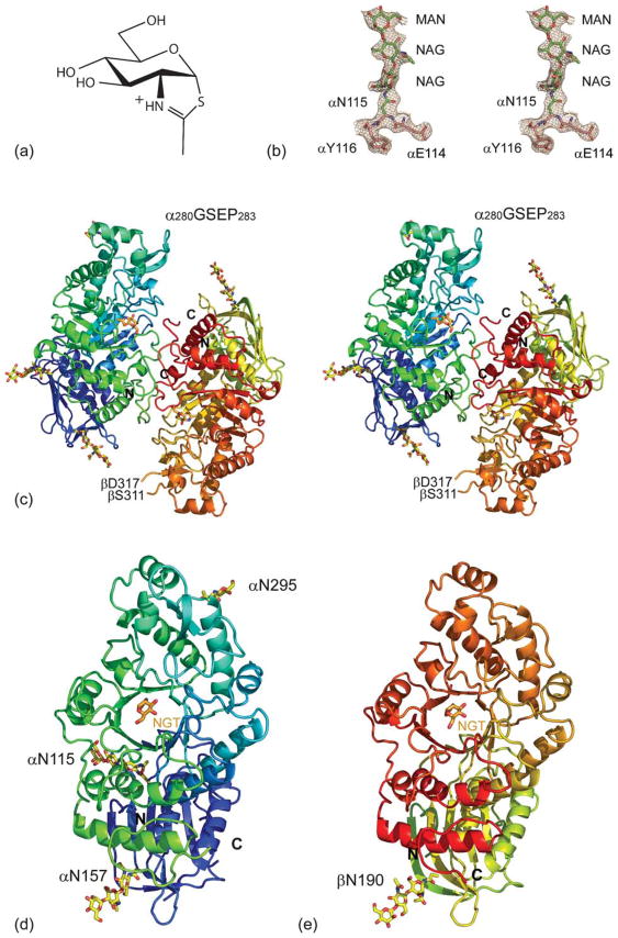
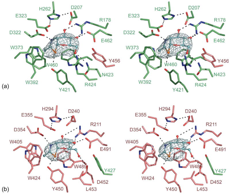
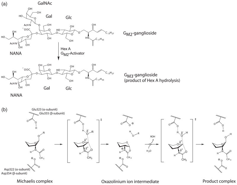
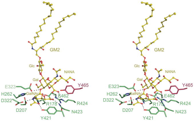
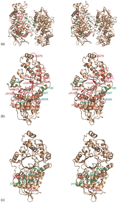
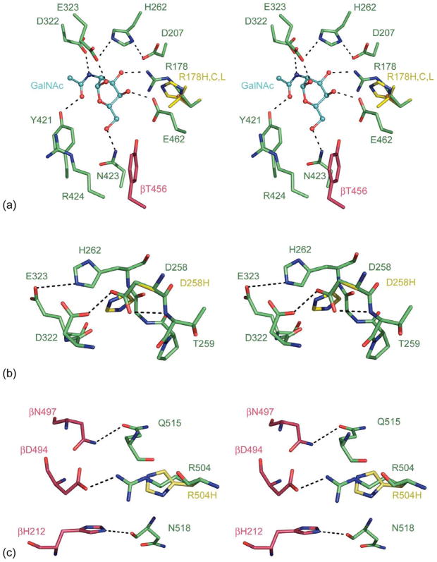
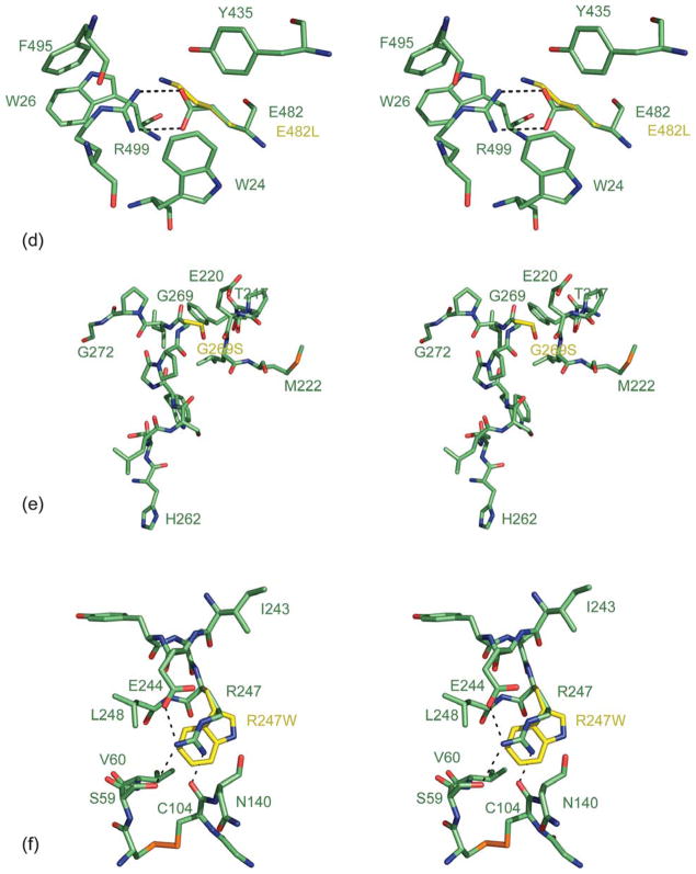
Lysosomal beta-hexosaminidase A (Hex A) is essential for the degradation of GM2 gangliosides in the central and peripheral nervous system. Accumulation of GM2 leads to severely debilitating neurodegeneration associated with Tay-Sachs disease (TSD), Sandoff disease (SD) and AB variant. Here, we present the X-ray crystallographic structure of Hex A to 2.8 A resolution and the structure of Hex A in complex with NAG-thiazoline, (NGT) to 3.25 A resolution. NGT, a mechanism-based inhibitor, has been shown to act as a chemical chaperone that, to some extent, prevents misfolding of a Hex A mutant associated with adult onset Tay Sachs disease and, as a result, increases the residual activity of Hex A to a level above the critical threshold for disease. The crystal structure of Hex A reveals an alphabeta heterodimer, with each subunit having a functional active site. Only the alpha-subunit active site can hydrolyze GM2 gangliosides due to a flexible loop structure that is removed post-translationally from beta, and to the presence of alphaAsn423 and alphaArg424. The loop structure is involved in binding the GM2 activator protein, while alphaArg424 is critical for binding the carboxylate group of the N-acetyl-neuraminic acid residue of GM2. The beta-subunit lacks these key residues and has betaAsp452 and betaLeu453 in their place; the beta-subunit therefore cleaves only neutral substrates efficiently. Mutations in the alpha-subunit, associated with TSD, and those in the beta-subunit, associated with SD are discussed. The effect of NGT binding in the active site of a mutant Hex A and its effect on protein function is discussed.


Figures

Figure 1
Figure 1
(a) Hex A structure. Chemical structure of NGT. (b) Stereo view of the 2FoFc map contoured at 1σ on residues αN114 to αY116, including glycosylation at αN115. (c) Stereo view of a ribbon representation for Hex A. The α-subunit N terminus color begins with dark blue and continues to light blue, and then ends with light green at its C terminus. The β-subunit N terminus begins with a greenish yellow color, changing to orange and ending in red at the C terminus. NGT, located at the face of the TIM barrel, is shown in orange. (d) The individual α-subunit and (e) β-subunit are represented as viewed from the dimer interface.
Figure 2
Figure 2
NGT bound in the active site of Hex A. (a) NGT (shown in blue) bound in the active site of the α-subunit (green) showing a minor contribution of βY456 from the β-subunit (pink). (b) NGT bound in the active site of the β-subunit (pink) showing a minor contribution of αY427 from the α-subunit (green). Unrefined FoFc density shown for NGT is contoured at 2.5σ.
Figure 3
Figure 3
Proposed catalytic mechanism for Hex A. (a) Hydrolysis of the GM2 ganglioside by Hex A results in the loss of GalNAc to produce a GM3 ganglioside. (b) Proposed catalytic mechanism for Hex A showing substrate-assisted catalysis. αGlu323 in the α-subunit and βGlu355 in the β-subunit act as the general base, while αAsp322 in the α-subunit and βAsp354 in the β-subunit act to orient the C2-acetamido group into position for nucleophilic attack and subsequently stabilizes the oxazolinium ion intermediate. The hydroxyl residues and C6 have been removed from the pyranose ring of the substrate for clarity. The exact positions for these groups have not been determined.
Figure 4
Figure 4
Model of GM2 docked onto the α-subunit active site of Hex A. A model of the GM2 ganglioside (yellow) was docked into the active site of the α-subunit of Hex A based on the model of GM2 bound to the α-subunit active site of Hex B. For clarity, only residues interacting with the sugar residues of GM2 are shown. GM2AP, which interacts with the acyl chains of the GM2 ganglioside, has also been removed. αArg424, a positively charged residue unique to the α-subunit of Hex A, is found within hydrogen bonding distance from the negatively charged carboxylate of the NANA group of GM2.
Figure 5
Figure 5
Known mutations of Hex A contributing to Tay-Sachs and Sandhoff disease. (a) Stereo view of a ribbon representation of Hex A (wheat), with residues known to disrupt Hex A activity: acute to sub-acute, red; chronic, green; asymptomatic, cyan. (b) A stereo view of the α-subunit of Hex A and residues associated with Tay-Sachs disease. (c) A stereo view of the β-subunit and residues associated with Sandhoff disease.
Figure 6
Figure 6
Model of Hex A mutants. (a) Stereo view representation of the Cα trace for wt-Hex A (α-subunit, green; β-subunit, pink) superimposed with the αArg178H,C,L Hex A substitutions (yellow). GalNAc (cyan) from a GM2 substrate has been docked into the active site. (b) Stereo view representation of the Cα trace for wt-Hex A (α-subunit, green) superimposed with the αAsp258His Hex A mutant (yellow). (c) A stereo view representation of the Cα trace for wt-Hex A (α-subunit, green; β-subunit, pink) superimposed with the αAsp258His substitution (yellow). (d) A stereo view representation of the Cα trace for wt-Hex A (α-subunit, green) superimposed with the αGlu482Lys Hex A substitutions (yellow). (e) A stereo view representation of the Cα trace for wt-Hex A (α-subunit, green) superimposed with the αGly269Ser Hex A substitutions (yellow). (f) A stereo view representation of the Cα trace for wt-Hex A (α-subunit, green) superimposed with the αArg247W Hex A mutant (yellow). Stick representations are shown for residues surrounding substitutions as well as other key residues described in the text.
Figure 6
Figure 6
Model of Hex A mutants. (a) Stereo view representation of the Cα trace for wt-Hex A (α-subunit, green; β-subunit, pink) superimposed with the αArg178H,C,L Hex A substitutions (yellow). GalNAc (cyan) from a GM2 substrate has been docked into the active site. (b) Stereo view representation of the Cα trace for wt-Hex A (α-subunit, green) superimposed with the αAsp258His Hex A mutant (yellow). (c) A stereo view representation of the Cα trace for wt-Hex A (α-subunit, green; β-subunit, pink) superimposed with the αAsp258His substitution (yellow). (d) A stereo view representation of the Cα trace for wt-Hex A (α-subunit, green) superimposed with the αGlu482Lys Hex A substitutions (yellow). (e) A stereo view representation of the Cα trace for wt-Hex A (α-subunit, green) superimposed with the αGly269Ser Hex A substitutions (yellow). (f) A stereo view representation of the Cα trace for wt-Hex A (α-subunit, green) superimposed with the αArg247W Hex A mutant (yellow). Stick representations are shown for residues surrounding substitutions as well as other key residues described in the text.

References

    1. Gravel RA, Clarke JTR, Kaback MM, Mahuran D, Sandoff K, Suzuki K. The GM2 gangliosidoses. In: Scriver CR, editor. The Metabolic and Molecular Basis of Inherited Diseases. McGraw-Hill: New York; 1995. pp. 2839–2879.
    1. Mahuran DJ. Biochemical consequences of mutations causing the GM2 gangliosidoses. Biochim Biophys Acta. 1999;1455:105–138. - PubMed
    1. Henrissat B, Davies G. Structural and sequence-based classification of glycoside hydrolases. Curr Opin Struct Biol. 1997;7:637–644. - PubMed
    1. Mahuran DJ. The GM2 activator protein, its roles as a co-factor in GM2 hydrolysis and as a general glycolipid transport protein. Biochim Biophys Acta. 1998;1393:1–18. - PubMed
    1. Kresse H, Fuchs W, Glossl J, Holtfrerich D, Gilberg W. Liberation of N-acetylgluco-samine-6-sulfate by human beta-N-acetylhexosaminidase A. J Biol Chem. 1981;256:12926–12932. - PubMed

Publication types

MeSH terms

Grants and funding