. 2006 Aug 1;91(3):996-1007.
doi: 10.1529/biophysj.105.079137. Epub 2006 May 12.

Secondary structure and Pd(II) coordination in S-layer proteins from Bacillus sphaericus studied by infrared and X-ray absorption spectroscopy

Affiliations

Secondary structure and Pd(II) coordination in S-layer proteins from Bacillus sphaericus studied by infrared and X-ray absorption spectroscopy

Karim Fahmy et al. Biophys J. .

Abstract

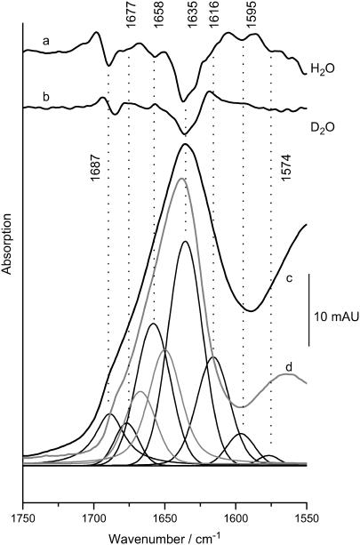
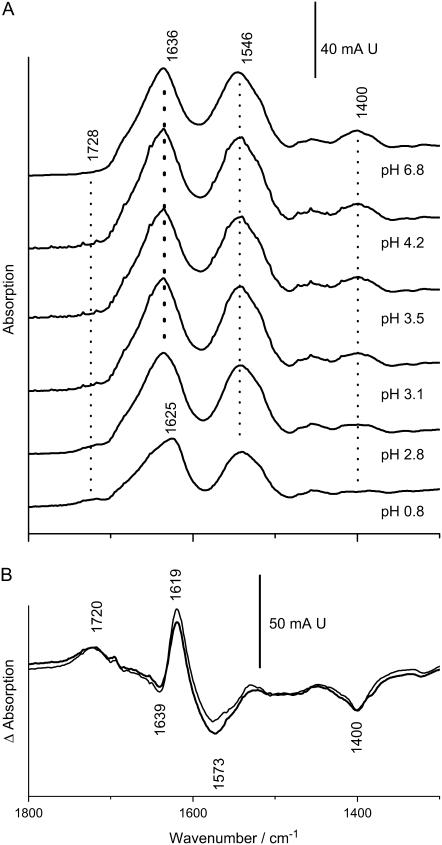
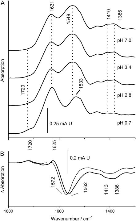
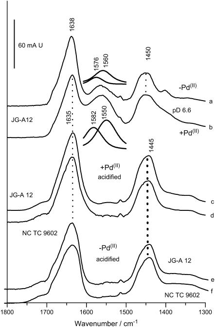
The S-layer of Bacillus sphaericus strain JG-A12, isolated from a uranium-mining site, exhibits a high metal-binding capacity, indicating that it may provide a protective function by preventing the cellular uptake of heavy metals and radionuclides. This property has allowed the use of this and other S-layers as self-assembling organic templates for the synthesis of nanosized heavy metal cluster arrays. However, little is known about the molecular basis of the metal-protein interactions and their impact on secondary structure. We have studied the secondary structure, protein stability, and Pd((II)) coordination in S-layers from the B. sphaericus strains JG-A12 and NCTC 9602 to elucidate the molecular basis of their biological function and of the metal nanocluster growth. Fourier transform infrared spectroscopy reveals similar secondary structures, containing approximately 35% beta-sheets and little helical structure. pH-induced infrared absorption changes of the side-chain carboxylates evidence a remarkably low pK < 3 in both strains and a structural stabilization when Pd((II)) is bound. The COO(-)-stretching absorptions reveal a predominant Pd((II)) coordination by chelation/bridging by Asp and Glu residues. This agrees with XANES and EXAFS data revealing oxygens as coordinating atoms to Pd((II)). The additional participation of nitrogen is assigned to side chains rather than to the peptide backbone. The topology of nitrogen- and carboxyl-bearing side chains appears to mediate heavy metal binding to the large number of Asp and Glu in both S-layers at particularly low pH as an adaptation to the environment from which the strain JG-A12 has been isolated. These side chains are thus prime targets for the design of engineered S-layer-based nanoclusters.


Figures

FIGURE 1
FIGURE 1
IR absorption in the amide spectral range of a suspension of S-layer from strain JG-A12. The structure-sensitive amide I mode (c) was approximated by fitted bands whose frequencies where determined from negative peaks in the second derivative of the spectrum measured in H2O (a). Original data and the sum of the fitted components are superimposed. The integral intensity of the bands above 1600 cm−1 was used to estimate the type and amount of secondary structure (summarized in Table 1). Disappearance of the shoulder at 1658 cm−1 in D2O (b) argues for a large contribution from turns and unordered structure rather than α-helices (52). (d) Absorption in D2O (shaded spectrum), where a better distinction between turns and random structure is achieved as evident from the larger separation of the two fitted bands (shaded) at 1667 and 1649 cm−1, respectively (for clarity only these two peaks in the amide I′ range are shown. Spectra are scaled to the 1635 cm−1 peak, which is barely affected in D2O as is typical of β-sheet absorption).
FIGURE 2
FIGURE 2
(A) pH-dependent IR-absorption of the S-layer of strain JG-A12. A suspension of S-layers (20–30 μL) was measured in attenuated total reflection on a Si-crystal (Bio-ATR-II) and was successively exposed to solutions of different pH (10 mM sodium phosphate) by dialysis. The protonation of carboxylic acids causes the reduction of the COO-stretching modes, which have lost half of their initial intensity at pH 3 (symmetric COO at 1400 cm−1). Irreversible denaturation is evidenced by the downshift of the amide I absorption at pH 0.7. (B) pH-induced absorption changes (spectra at pH 0.8 minus spectra at pH 7) for S-layers of JG-A12 (thick line) and NCTC 9602 (thin line) visualize both the carboxylate and amide I absorption change in the pH 7 to pH 0.7 interval.
FIGURE 3
FIGURE 3
(A) pH-dependent IR-absorption of Pd(II)-bound S-layer of strain JG-A12. (B) pH-induced absorption changes in S-layers from strain JG-A12 (thick line) and NCTC 9602 (thin line) calculated as in Fig. 2 show the carboxylate absorption changes, whereas amide absorption changes are largely blocked. The asymmetric COO-stretching modes are enhanced versus the native proteins and their average frequency is downshifted by 10 cm−1 in JG-A12, whereas it is not altered in NCTC 9602. Experimental conditions as in legend to Fig. 2.
FIGURE 4
FIGURE 4
SDS-gelelectrophoresis. (A) The S-layer proteins of B. sphaericus JG-A12 (lane 1) and NCTC 9602 (lane 2) were digested with Glu-C and separated using a 12.5% gel. M, marker. (B) Native and Pd(II)-complexed S-layer proteins of B. sphaericus JG-A12 were digested with Glu-C and analyzed using a 10% gel. Lane 1: purified S-layer protein. Lane 2: Glu-C. Lane 3: purified S-layer protein with complexed Pd(II) after digestion with Glu-C. Lane 4: S-layer protein with complexed Pd(II); M, marker.
FIGURE 5
FIGURE 5
Pd K-edge XANES region of EXAFS spectra of Pd-loaded S-layer protein of B. sphaericus JG-A12 and reference compounds (Pd foil, PdO, Na2PdCl4, [Pd(NH3)4]Cl2, and PdCl2).
FIGURE 6
FIGURE 6
Pd K-edge k3-weighted EXAFS spectra (left) and the corresponding FT (right) of the Pd reference compounds.
FIGURE 7
FIGURE 7
Pd K-edge k3-weighted EXAFS spectra (left) and the corresponding FT (right) of the Pd complexes formed by S-layer of B. sphaericus JG-A12 according to model a lower traces and model b upper traces.
FIGURE 8
FIGURE 8
FT of the EXAFS spectra of Pd-treated S-layers at pH 2.0 and pH 3.1. Note the constant ratio of the amplitudes of the peak A and peak B indicating that the latter may be a side lobe of peak A. See text for details.
FIGURE 9
FIGURE 9
FTIR spectra of S-layers of B. sphaericus strain JG-A12 and NCTC 9602 measured in D2O. (a) Spectrum of JG-A12 at pH 7, metal free. (b) Spectrum of JG-A12 at pH 7, Pd(II) bound. Note the broadening of the 1400 cm−1 absorption (symmetric COO stretching) and the increase of the ∼1560 cm−1 absorption (antisymmetric COO- stretching) as compared to a. The antisymmetric COO-stretching modes where fitted by two components absorbing at 1576 and 1560 cm−1 (inset). Besides the strong absorption enhancement, the salient effect of Pd(II) binding is the 10 cm−1 downshift of the 1560 cm−1 absorption, whereas the upshift of the high frequency part is less pronounced. (c) Absorption of JG-A12 in the Pd(II)-bound state acidified with DCl. (d) Absorption of NCTC 9602 in the Pd(II)-bound state acidified with DCl. Note the absence of residual absorption in the 1560–1570 cm−1 range and the lack of pH sensitivity in the amide II′ mode (1450 cm−1) in c and d as compared to a and b. (e) Absorption of JG-A12 in the metal-free state acidified with DCl. (f) Absorption of NCTC 9602 in the metal-free state acidified with DCl. Note the correspondence of the amide II′ modes in c and e and in d and f.

References

    1. Sleytr, U. B., and M. Sara. 1997. Bacterial and archaeal S-layer proteins: structure-function relationships and their biotechnological applications. Trends Biotechnol. 15:20–26. - PubMed
    1. Beveridge, T. J. 1994. Bacterial S-layers. Curr. Opin. Struct. Biol. 4:204–212.
    1. Dieluweit, S., D. Pum, and U. B. Sleytr. 1998. Formation of a gold superlattice on an S-layer with square lattice symmetry. Supramolecular Sci. 5:15–19.
    1. Engelhardt, H., and J. Peters. 1998. Structural research on surface layers: a focus on stability, surface layer homology domains, and surface layer-cell wall interactions. J. Struct. Biol. 124:276–302. - PubMed
    1. Selenska-Pobell, S., P. Panak, V. Miteva, G. Bernhard, and H. Nitsche. 1999. Selective accumulation of heavy metals by three indigenous Bacillus strains, B. cereus, B. megaterium and B. sphaericus, from drain waters of a uranium waste pile. FEMS Microbiol. Ecol. 29:59–67.

Publication types

MeSH terms