

doi: 10.1529/biophysj.106.085100.
Epub 2006 May 12.
Tryptophan rotamers as evidenced by X-ray, fluorescence lifetimes, and molecular dynamics modeling
Affiliations
- PMID: 16698786
- PMCID: PMC1563760
- DOI: 10.1529/biophysj.106.085100
Item in Clipboard
Tryptophan rotamers as evidenced by X-ray, fluorescence lifetimes, and molecular dynamics modeling
Biophys J.
.
Abstract
We investigated the native-state dynamics of the Bacillus caldolyticus cold-shock protein mutant Bc-Csp L66E, using fluorescence and appropriate molecular dynamics methods. Two fluorescence lifetimes were found, the amplitudes of which agree very well with tryptophan rotamer populations, obtained from parallel tempering calculations. Rotamer lifetimes were predicted by transition-state theory from high-temperature simulations. Transition pathways were extracted from the transition rates between individual rotameric states. The molecular dynamics also reveal the loop fluctuations in the native state.
Figures
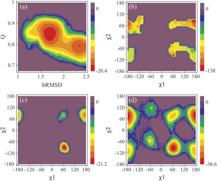
Energy contour maps in units of kJ mol−1. (a) Free energy as a function of Q and bRMSD, from PT. The highest bRMSD well corresponds with a loss of the 310-helix structure. (b) Potential energy in function of Trp8 χ1 and χ2, from DEE. The energies are calculated as the total nonbonded energies Etotnb for all structures with a negative Etotnb. Free energy in function of Trp8 χ1 and χ2 from (c) PT and (d) 500 K MD. The relative free energy ΔG between two states i and j with relative populations Pi and Pj is calculated as  , where R denotes the universal gas constant.
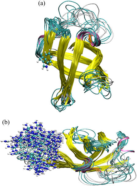
Superposition of 15 native-state structures of 1HZB from PT. The structures were carefully chosen to represent the occupied conformational space. The color scheme is as follows: 310-helix (mauve), extended β-strand (yellow), isolated β-bridge (orange), hydrogen-bonded turn (cyan), and coil (white). To these ribbon models were added in ball-and-stick models the side chains of (a) Trp8 in the p−90° state, and (b) 118 Arg56 conformations.
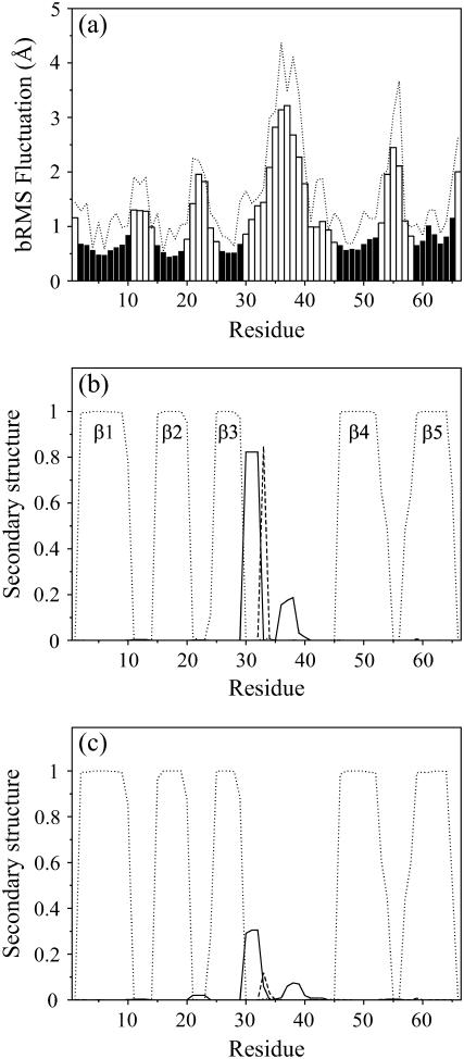
(a) bRMS fluctuation (bars) and total RMS fluctuation (dotted line) as a function of the residue number, from PT. The black bars indicate β-sheet residues. (b and c) Secondary-structure content in function of residue number for states belonging to (b) the low-bRMSD free-energy well and (c) the high-bRMSD free-energy well (see Fig. 1 a). Curves represent the 310-helix (solid lines), isolated β-bridge (dashed lines), and extended β-strand (dotted lines).
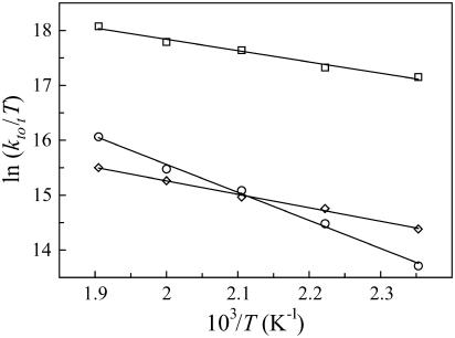
Eyring plots from the high-temperature dynamics for the three most stable rotamers, based on the total transition rate constants ktot,i (T). p−90° (○), t80° (⋄), and p80° (□).
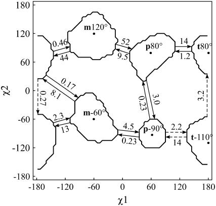
χ1 × χ2 map of the six stable rotamers and a schematic representation of the observed transitions at 500 K, with transition rates given in ns−1. The dashed arrows indicate the reaction path between p−90° and t80°. The contour lines enclose 95% of the population of each rotamer.
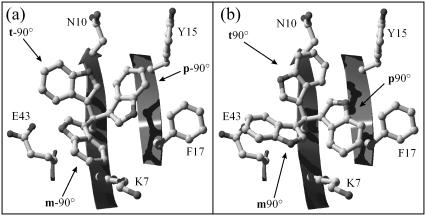
Tryptophan rotamers in Bc-Csp 1HZB. Superposition of the six Trp8 rotamers in their ideal positions and the surface-exposed residues within a 5-Å radius of Trp8. (a) m−90°, p−90°, and t−90°. (b) m90°, p90°, and t90°.
References
-
- Osterberg, F., G. M. Morris, M. F. Sanner, A. J. Olson, and D. S. Goodsell. 2002. Automated docking to multiple target structures: incorporation of protein mobility and structural water heterogeneity in AutoDock. Proteins. 46:34–40. - PubMed
-
- Stapley, B. J., and A. J. Doig. 1997. Free energies of amino acid side-chain rotamers in α-helices, β-sheets and α-helix N-caps. J. Mol. Biol. 272:456–464. - PubMed
-
- Lindorff-Larsen, K., P. Rogen, E. Paci, M. Vendruscolo, and C. M. Dobson. 2005. Protein folding and the organization of the protein topology universe. Trends Biochem. Sci. 30:13–19. - PubMed
Publication types
MeSH terms
Substances
LinkOut - more resources
Full Text Sources
