. 2006 Jul;141(3):851-7.
doi: 10.1104/pp.106.081091. Epub 2006 May 12.

Early steps in the biosynthesis of NAD in Arabidopsis start with aspartate and occur in the plastid

Affiliations

Early steps in the biosynthesis of NAD in Arabidopsis start with aspartate and occur in the plastid

Akira Katoh et al. Plant Physiol. 2006 Jul.

Abstract

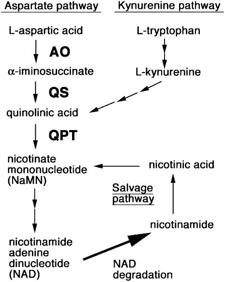
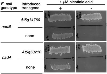
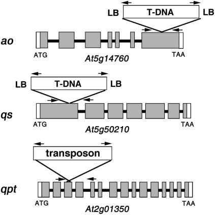
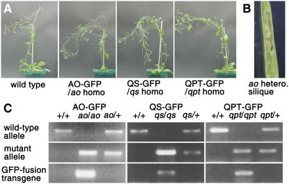
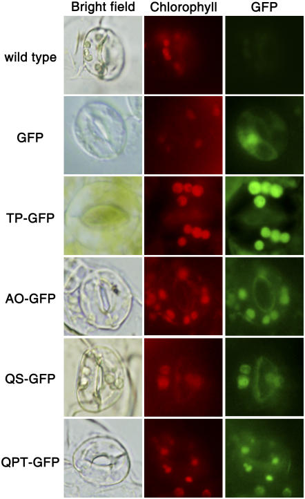
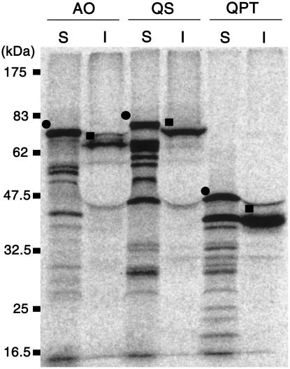
NAD is a ubiquitous coenzyme involved in oxidation-reduction reactions and is synthesized by way of quinolinate. Animals and some bacteria synthesize quinolinate from tryptophan, whereas other bacteria synthesize quinolinate from aspartate (Asp) using L-Asp oxidase and quinolinate synthase. We show here that Arabidopsis (Arabidopsis thaliana) uses the Asp-to-quinolinate pathway. The Arabidopsis L-Asp oxidase or quinolinate synthase gene complemented the Escherichia coli mutant defective in the corresponding gene, and T-DNA-based disruption of either of these genes, as well as of the gene coding for the enzyme quinolinate phosphoribosyltransferase, was embryo lethal. An analysis of functional green fluorescent protein-fused constructs and in vitro assays of uptake into isolated chloroplasts demonstrated that these three enzymes are located in the plastid.


Figures

Figure 1.
Figure 1.
Alternate pathways for NAD biosynthesis. Some bacteria, such as E. coli, synthesize quinolinic acid from Asp using AO and QS, whereas animals, fungi, and other bacteria produce quinolinate from Trp via kynurenine. Quinolinate is converted to NAD in three steps, with the first step catalyzed by QPT. In some biochemical reactions, NAD is metabolized to nicotinamide, which is recycled back to NaMN via a salvage pathway.
Figure 2.
Figure 2.
Complementation of E. coli AO and QS mutants with the Arabidopsis homologs. E. coli strains nadB and nadA, which were deficient in AO and QS, respectively, were transformed with the vectors that expressed the Arabidopsis homolog of AO (At5g14760) or QS (At5g50210), or with an empty vector (none). Transformed bacteria were cultured on minimal medium containing 1 mm isopropylthio-β-galactoside and 50 μg/mL ampicilin in the presence (+) or absence (−) of 1 μm nicotinic acid.
Figure 3.
Figure 3.
Gene-disrupted alleles of Arabidopsis AO, QS, and QPT. The positions of the T-DNA and dissociation transposon insertion in AO (At5g14760), QS (At5g50210), and QPT (At2g01350) are schematically shown. In qpt, the transposon's insertion generated a small tandem duplication of a part of the third exon. Gray and white boxes represent protein-coding regions and untranslated flanking sequences, whereas thick lines represent introns. Approximate positions of PCR primers are indicated by arrows. LB, Left border of T-DNA.
Figure 4.
Figure 4.
Functional complementation of homozygous null mutants with the constitutive expression of AO-GFP, QS-GFP, or QPT-GFP. A, Two-month-old adult wild-type plants (ecotype Columbia) and homozygous mutant plants expressing GFP-fusion transgenes. B, A young silique of a self-pollinated heterozygous ao mutant plant. Development was affected in about 25% of seeds. C, Genomic PCR analysis. The wild type (+/+), heterozygotes (mutant allele/+), and homozygotes (mutant allele/mutant allele) expressing relevant GFP-fusion transgenes were analyzed for the presence of wild-type alleles, T-DNA/transposon insertion mutant alleles, and GFP-fusion transgenes.
Figure 5.
Figure 5.
GFP-fused proteins of AO, QS, and QPT are localized to the chloroplasts of Arabidopsis guard cells. Chlorophyll autofluorescence (red) and GFP fluorescence (green) in guard cells were simultaneously observed. Bright-field images on the right show the shape of guard cells. Unfused GFP was found in the cytoplasm and nucleus, whereas TP-GFP (plastid-targeted positive control), AO-GFP, QS-GFP, and QPT-GFP were all found in the chloroplasts.
Figure 6.
Figure 6.
Import of protein into chloroplasts in vitro. After the translated radiolabeled substrates (S) of AO, QS, or QPT were incubated with isolated pea chloroplasts, imported proteins in the chloroplasts (I) were analyzed. Precursor and mature forms of AO, QS, and QPT are, respectively, indicated by black circles and black squares in the top left of the bands. The size of Mr markers is shown on the left.

References

    1. Ashihara H, Stasolla C, Yin Y, Loukanina N, Thorpe TA (2005) De novo and salvage biosynthetic pathways of pyridine nucleotides and nicotinic acid conjugates in cultured plant cells. Plant Sci 169: 107–114
    1. Baginsky S, Siddique A, Gruissem W (2004) Proteome analysis of tobacco bright yellow-2 (BY-2) cell culture plastids as a model for undifferentiated heterotrophic plastids. J Proteome Res 3: 1128–1137 - PubMed
    1. Bruce BD, Perry S, Froehlich J, Keegstra K (1994) In vitro import of protein into chloroplasts. In SB Gelvin, RB Schilperoort, eds, Plant Molecular Biology Manual, Ed 2, Vol J1. Kluwer Academic Publishers, Boston, pp 1–15
    1. Chen D, Steele AD, Lindquist S, Guarente L (2005) Increase in activity during calorie restriction requires Sirt1. Science 310: 1641. - PubMed
    1. Chiu W, Niwa Y, Zeng W, Hirano T, Kobayashi H, Sheen J (1996) Engineered GFP as a vital reporter in plants. Curr Biol 6: 325–330 - PubMed

Publication types

MeSH terms