. 2006 Jul;141(3):898-909.
doi: 10.1104/pp.106.080945. Epub 2006 May 12.

Surface position, not signaling from surrounding maternal tissues, specifies aleurone epidermal cell fate in maize

Affiliations

Surface position, not signaling from surrounding maternal tissues, specifies aleurone epidermal cell fate in maize

Darren Fred Gruis et al. Plant Physiol. 2006 Jul.

Erratum in

  • Plant Physiol. 2006 Dec;142(4):1771

Abstract

Maize (Zea mays) endosperm consists of an epidermal-like surface layer of aleurone cells, an underlying body of starchy endosperm cells, and a basal layer of transfer cells. To determine whether surrounding maternal tissues perform a role in specifying endosperm cell fates, a maize endosperm organ culture technique was established whereby the developing endosperm is completely removed from surrounding maternal tissues. Using cell type-specific fluorescence markers, we show that aleurone cell fate specification occurs exclusively in response to surface position and does not require specific, continued maternal signal input. The starchy endosperm and aleurone cell fates are freely interchangeable throughout the lifespan of the endosperm, with internalized aleurone cells converting to starchy endosperm cells and with starchy endosperm cells that become positioned at the surface converting to aleurone cells. In contrast to aleurone and starchy endosperm cells, transfer cells fail to develop in in vitro-grown endosperm, supporting earlier indications that maternal tissue interaction is required to fully differentiate this cell type. Several parameters confirm that the maize endosperm organ cultures described herein retain the main developmental features of in planta endosperm, including fidelity of aleurone mutant phenotypes, temporal and spatial control of cell type-specific fluorescent markers, specificity of cell type transcripts, and control of mitotic cell divisions.


Figures
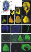
Figure 1.
Figure 1.
Anatomy and structure of in planta- and in vitro-grown maize endosperms. A, Cartoon showing three endosperm cell types in the colors of the transgenic maize triple-marker line. al, Aleurone; e, embryo; se, starchy endosperm; tc, transfer cells (same section plane as in B–D). B, Transverse section of 6-DAP ovule with fertilized embryo sac (arrow) embedded in nucellus parenchyma cells. C, Transverse section of 10-DAP grain. D, Transverse section of 15-DAP grain. E, Intact unstained in vitro endosperms harvested at 6 DAP and grown in vitro for various time intervals. F, Six-DAP endosperms grown for 2 DIV. G, Six-DAP endosperms grown for 4 DIV. H, Six-DAP endosperms grown for 10 DIV. In planta- or in vitro-grown endosperms are from the triple-marker line. I, γ-Zein:AmCyan expression in the starchy endosperm of 12-DAP in planta endosperms. J, γ-Zein:AmCyan in starchy endosperm cells of 6-DIV in vitro-grown endosperms. K, Ltp2:ZsYellow in the aleurone layer of 12-DAP in planta endosperms. L, Ltp2:ZsYellow in the surface aleurone cell layer of 6-DIV in vitro endosperms. M to P, Expression of Ltp2:ZsYellow in the surface layer of aleurone cells of in vitro-grown endosperms harvested at 6 DAP (M) and grown for 2 (N), 4 (O), and 6 DIV (P). Q, End1:DsRed expression in the transfer cell layer of 12-DAP in planta endosperms. R, View of the basal exterior surface of 6-DIV in vitro-grown endosperms from the triple-marker line, with the area where transfer cells form in planta lacking red fluorescences shown in black and adjoining aleurone cells with ZsYellow fluorescence. tca, Transfer cell area.
Figure 2.
Figure 2.
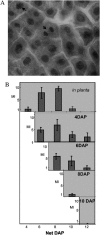
Mitotic activity in the surface layer of in planta- and in vitro-grown endosperms. A, Light micrograph of aceto-orcein-stained aleurone layer at 6 DAP showing cells in mitosis (arrows) and interphase. B, Top, Mitotic index in endosperms fixed in the interval from 4 to 12 DAP. B, Remaining images, Mitotic index of in vitro-grown endosperms harvested at 4, 6, 8, and 10 DAP and grown in in vitro culture for 8, 6, 4, and 2 d, respectively, and examined at 2-d intervals during culture. y axis indicates mitotic index (MI) and x axis indicates net DAP.
Figure 3.
Figure 3.
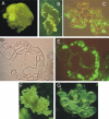
Endosperms from suspension cultures grown in medium with 3% Suc contain a high proportion of nondifferentiated cells and lack internal organization. A, Endosperms grown in vitro for 15 DIV showing growth of endosperm cells on the basal surface. B, Light micrograph of callus at 45 DIV with firm surface. C, Transverse section of callus in B showing irregular pattern of Ltp2:ZsYellow fluorescence in internal cells intermingled with nonfluorescing high-vacuolated cells. D, Phase contrast micrograph of spherical structure from B with aleurone-like layer surface cells and highly vacuolated internal cells. E, Same as in D shown in epifluorescence and visible light. F, Loosely organized callus of unspecialized cells. G, Higher magnification of cells in callus type shown in F.
Figure 4.
Figure 4.
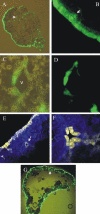
Expression of the Ltp2:ZsYellow fluorescence marker confirms that cells in surface position assume aleurone cell fate. A, Mixed light and epifluorescence micrograph of transverse section of 6-DIV in vitro endosperms showing ZsYellow fluorescence in the surface layer as well as in some areas in the interior of the starchy endosperm (one example indicated by arrow). B, Mixed light and epifluorescence micrograph of the peripheral endosperm layer of section shown in A with strong fluorescence in the outer layer and weaker fluorescence in the second cell layer. Arrow identifies a cell in the second layer that shows strong fluorescence. C, Mixed phase contrast and epifluorescence micrograph showing Ltp2:ZsYellow fluorescence in internal cells adjacent to a void (V) in the endosperm identified by an arrow in A. D, Same as in C shown in epifluorescence microscopy. E, Laser confocal microscopy image showing the periphery of in vitro-grown endosperms at 15 DIV showing Ltp2:ZsYellow in aleurone cells and γ-Zein:AmCyan in starchy endosperms. Arrow identifies aleurone cells around an internal void. F, Higher magnification of interior aleurone cells in E. G, Transverse section of in vitro-grown endosperms at 15 DIV, where the interior starchy endosperms failed to develop showing partial aleurone layers on the interior side of the endosperm (arrow).
Figure 5.
Figure 5.
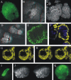
Development of mini-endosperms in in vitro-cultured endosperms. A, Epifluorescence micrograph showing surface view of 15-DIV triple-marker line endosperms with numerous mini-endosperms fluorescing strongly with ZsYellow. B, A 3-D confocal image reconstruction of aleurone cells on the surface of 10-DIV in vitro-grown endosperms showing sectors with different intensities of ZsYellow fluorescence (red-green stereo anaglyphs required to see stereo effect). C, A 3-D confocal image reconstruction of early mini-endosperms at 12 DIV. D, A 3-D confocal image reconstruction of fully developed mini-endosperms at 15 DIV. E, Epifluorescence micrograph of transverse section of 15-DIV in vitro endosperms similar to A showing abundant mini-endosperm covered with Ltp2:ZsYellow positive aleurone cells. F, Confocal image of transverse section of mini-endosperms showing surface layer of aleurone cells expressing Ltp2:ZsYellow and interior starchy endosperm cells expressing γ-Zein:AmCyan. G and H, Side-by-side stereogram confocal micrographs showing 3-D reconstructions of surface layer of mini-endosperms fluorescing with ZsYellow. G, Outside view from the top. H, Interior view from the base and up. I to K, Sectors and mini-endosperm formed after particle bombardment transformation of young surface cells with a Ltp2∷ZsYellow construct. I, Sector of Ltp2:ZsYellow positive cells in 10-DIV wild-type in vitro endosperms shown in epifluorescence microscopy. J, A 3-D reconstruction of Ltp2:ZsYellow positive sector shown in side-by-side stereogram confocal image. K, Complete mini-endosperm formed after bombardment shown in epifluorescence microscopy.
Figure 6.
Figure 6.
dek1 and sal1-2 mutant endosperms grown in vitro have similar phenotypes as in planta. A, Phase contrast micrograph of typical dek1 endosperms cultured in vitro. B, Same as in A shown in epifluorescence showing a surface layer of cuboidal cells. Arrow identifies peripheral cell that expresses the Ltp2:ZsYellow marker. C and D, Enlargement of the Ltp2:ZsYellow positive cell of B in phase contrast (C) and in epifluorescence microscopy (D). E, Phase contrast micrograph of transverse section of a typical dek1 mutant endosperm with surface layer of aleurone-like cells and mini-endosperms lacking aleurone cells. Arrow identifies area with three Ltp2:ZsYellow positive cells of section E shown in F and G at higher magnification. F, Phase contrast and epifluorescence micrograph of peripheral area identified by arrow in E. G, Phase contrast micrograph of area shown in F. H, Autofluorescence image showing enlargement of mini-endosperms in section shown in E. Arrow identifies mini-endosperms with organized layers of surface cells. I, Transverse section of sal1-2 mutant endosperms with double layers of aleurone cells (identified by arrows) from in vitro culture.

References

    1. Ahn J-W, Kim M, Lim JH, Kim G-T, Pai H-S (2004) Phytocalpain controls the proliferation and differentiation fates of cells in plant organ development. Plant J 38: 969–981 - PubMed
    1. Becraft PW, Asuncion-Crabb Y (2000) Positional cues specify and maintain aleurone cell fate in maize endosperm development. Development 127: 4039–4048 - PubMed
    1. Becraft PW, Stinard PS, McCarty D (1996) CRINKLY4: a TNFR-like receptor kinase involved in maize epidermal differentiation. Science 273: 1406–1409 - PubMed
    1. Cao X, Li K, Suh SG, Guo T, Becraft PW (2005) Molecular analysis of the CRINKLY4 gene family in Arabidopsis thaliana. Planta 220: 645–657 - PubMed
    1. Castro MS, Fontes W, Morhy L, Bloch CJ (1996) Complete amino acid sequences of two gamma-thionins from maize (Zea mays L.) seeds. Protein Pept Lett 3: 267–274

LinkOut - more resources