. 2006 Jul;141(3):957-65.
doi: 10.1104/pp.106.079319. Epub 2006 May 12.

NUCLEAR FUSION DEFECTIVE1 encodes the Arabidopsis RPL21M protein and is required for karyogamy during female gametophyte development and fertilization

Affiliations

NUCLEAR FUSION DEFECTIVE1 encodes the Arabidopsis RPL21M protein and is required for karyogamy during female gametophyte development and fertilization

Michael F Portereiko et al. Plant Physiol. 2006 Jul.

Abstract

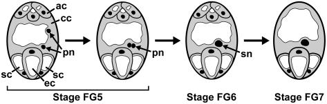
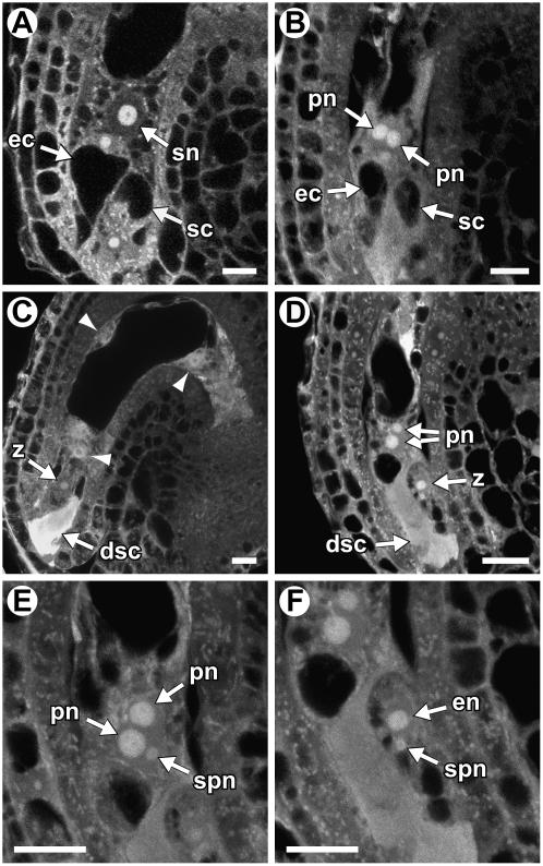
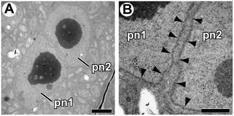
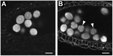
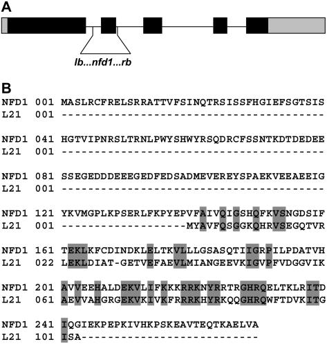
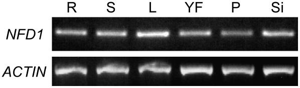
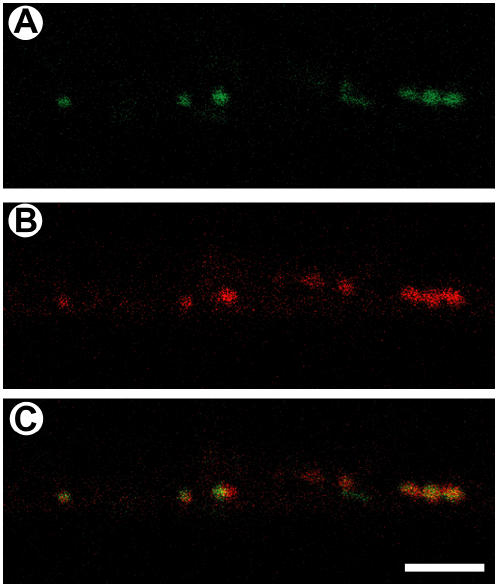
Karyogamy, or nuclear fusion, is essential for sexual reproduction. In angiosperms, karyogamy occurs three times: twice during double fertilization of the egg cell and the central cell and once during female gametophyte development when the two polar nuclei fuse to form the diploid central cell nucleus. The molecular mechanisms controlling karyogamy are poorly understood. We have identified nine female gametophyte mutants in Arabidopsis (Arabidopsis thaliana), nuclear fusion defective1 (nfd1) to nfd9, that are defective in fusion of the polar nuclei. In the nfd1 to nfd6 mutants, failure of fusion of the polar nuclei is the only defect detected during megagametogenesis. nfd1 is also affected in karyogamy during double fertilization. Using transmission electron microscopy, we showed that nfd1 nuclei fail to undergo fusion of the outer nuclear membranes. nfd1 contains a T-DNA insertion in RPL21M that is predicted to encode the mitochondrial 50S ribosomal subunit L21, and a wild-type copy of this gene rescues the mutant phenotype. Consistent with the predicted function of this gene, an NFD1-green fluorescent protein fusion protein localizes to mitochondria and the NFD1/RPL21M gene is expressed throughout the plant. The nfd3, nfd4, nfd5, and nfd6 mutants also contain T-DNA insertions in genes predicted to encode proteins that localize to mitochondria, suggesting a role for this organelle in nuclear fusion.


Figures

Figure 1.
Figure 1.
Karyogamy during female gametophyte development in Arabidopsis. Female gametophyte developmental stages: The megaspore (stage FG1; not shown) undergoes three rounds of mitosis and cellularization and results in a seven-celled, eight-nucleate female gametophyte (stage FG5). The central cell inherits two nuclei (polar nuclei), which migrate toward the female gametophyte's center and fuse to form the secondary nucleus (stage FG6). Finally, the antipodal cells degenerate, producing a mature female gametophyte (stage FG7). ac, Antipodal cells; cc, central cell; ec, egg cell; pn, polar nuclei; sc, synergid cell; sn, secondary nucleus. In these images, nuclei are black, cytoplasm is gray, and vacuoles are white.
Figure 2.
Figure 2.
CLSM analysis of wild-type and nfd1 female gametophytes. A and B, Wild type (A) and nfd1 (B) female gametophytes at the terminal developmental stage (stage FG7). In wild type (A), the polar nuclei have fused to form the diploid secondary nucleus. A wild-type egg cell (A) contains a single, large vacuole and has a nucleus at the chalazal pole. In nfd1 (B), the polar nuclei remain unfused. C to F, Wild-type (C) and nfd1 (D–F) seeds at 18 h after pollination. The wild-type seed (C) contains a degenerating synergid cell, a zygote, and four endosperm nuclei (arrowheads; only three endosperm nuclei are in this focal plane). A wild-type zygote (C) is relatively round (in comparison to the egg cell), contains many small vacuoles, and has a centrally located nucleus. nfd1 (D) contains a degenerated synergid cell, a zygote, two unfused polar nuclei, and no endosperm nuclei. E and F, Magnified images from the same ovule as in D. The image in E is of a different focal plane than that in D. The image in F is of the same focal plane as that in D. In E, the polar nuclei and the sperm nucleus are visible within the central cell. In F, the egg cell nucleus and the sperm nucleus are visible within the zygote. In these images, cytoplasm is gray, vacuoles are black, and nucleoli are white. dsc, Degenerating synergid cell; ec, egg cell; en, egg cell nucleus; pn, polar nucleus; sc, synergid cell; sn, secondary nucleus; spn, sperm nucleus; z, zygote. Scale bars = 10 μm.
Figure 3.
Figure 3.
TEM analysis of the unfused polar nuclei in nfd1 female gametophytes. A and B, Electron micrographs of the central cell of female gametophytes at the terminal developmental stage (stage FG7). The nucleoli (dark gray) are surrounded by a nucleoplasm (light gray) and a nuclear envelope (arrowheads). Scale bars = 3 μm (A) and 1 μm (B). pn, Polar nucleus.
Figure 4.
Figure 4.
CLSM analysis of pollen from wild type and nfd1/NFD1. A, Pollen from wild-type anthers. The pollen grains are round. B, Pollen from nfd1/NFD1 anthers. Both round and collapsed pollen grains are present. Arrowheads indicate two of the collapsed pollen grains in this image. Scale bars = 20 μm (A and B).
Figure 5.
Figure 5.
NFD1 gene structure and protein alignment. A, NFD1 gene structure. Black boxes represent coding sequence, gray boxes represent UTRs, and the horizontal line represents intron sequence. The vertical lines represent the T-DNA insertion sites in nfd1. B, Comparison of the amino acid sequence between NFD1 and the L21p protein in E. coli. Conserved amino acids are in gray.
Figure 6.
Figure 6.
RT-PCR analysis of NFD1 expression. Shown is RT-PCR analysis of NFD1 in roots (R), inflorescence stems (S), leaves (L), young flowers (YF), pistils (P), and siliques (Si). Expression of ACTIN was used as a control.
Figure 7.
Figure 7.
NFD1-GFP localization. Shown are CLSM images of a MitoTracker-stained root from a transgenic plant expressing NFD1-GFP. A, GFP fluorescence. B, MitoTracker red fluorescence. C, Merge of A and B. Scale bar = 5 μm.

Similar articles

Cited by

References

    1. Alexander RW, Cooperman BS (1998) Ribosomal proteins neighboring 23 S rRNA nucleotides 803-811 within the 50 S subunit. Biochemistry 37: 1714–1721 - PubMed
    1. Beh CT, Brizzio V, Rose MD (1997) KAR5 encodes a novel pheromone-inducible protein required for homotypic nuclear fusion. J Cell Biol 139: 1063–1076 - PMC - PubMed
    1. Boisnard-Lorig C, Colon-Carmona A, Bauch M, Hodge S, Doerner P, Bancharel E, Dumas C, Haseloff J, Berger F (2001) Dynamic analyses of the expression of the HISTONE::YFP fusion protein in Arabidopsis show that syncytial endosperm is divided in mitotic domains. Plant Cell 13: 495–509 - PMC - PubMed
    1. Brizzio V, Khalfan W, Huddler D, Beh CT, Andersen SS, Latterich M, Rose MD (1999) Genetic interactions between KAR7/SEC71, KAR8/JEM1, KAR5, and KAR2 during nuclear fusion in Saccharomyces cerevisiae. Mol Biol Cell 10: 609–626 - PMC - PubMed
    1. Choi Y, Gehring M, Johnson L, Hannon M, Harada JJ, Goldberg RB, Jacobsen SE, Fischer RL (2002) DEMETER, a DNA glycosylase domain protein, is required for endosperm gene imprinting and seed viability in Arabidopsis. Cell 110: 33–42 - PubMed

Publication types

Associated data