. 2006 May 23;103(21):8131-6.
doi: 10.1073/pnas.0602498103. Epub 2006 May 12.

Hsk1 kinase is required for induction of meiotic dsDNA breaks without involving checkpoint kinases in fission yeast

Affiliations

Hsk1 kinase is required for induction of meiotic dsDNA breaks without involving checkpoint kinases in fission yeast

Keiko Ogino et al. Proc Natl Acad Sci U S A. .

Abstract

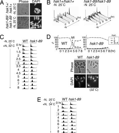
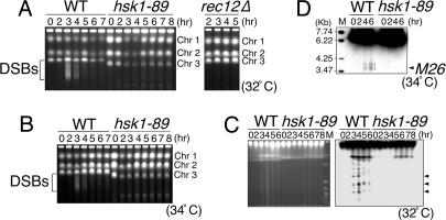
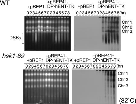
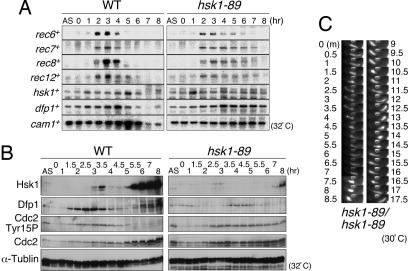
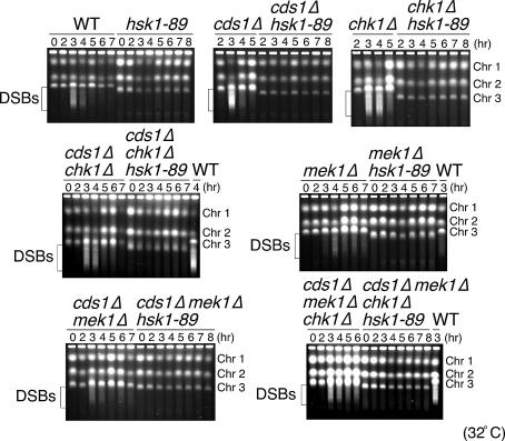
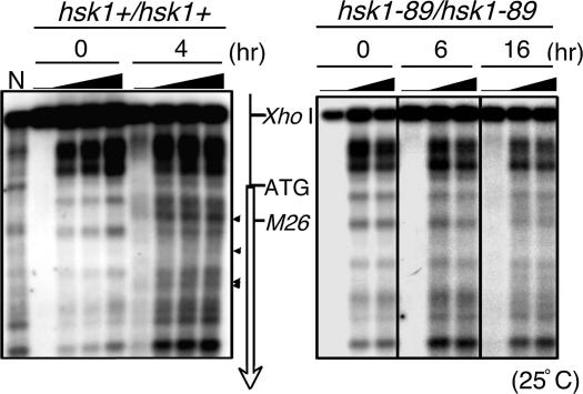
Cdc7 kinase, conserved through evolution, is known to be essential for mitotic DNA replication. The role of Cdc7 in meiotic recombination was suggested in Saccharomyces cerevisiae, but its precise role has not been addressed. Here, we report that Hsk1, the Cdc7-related kinase in Schizosaccharomyces pombe, plays a crucial role during meiosis. In a hsk1 temperature-sensitive strain (hsk1-89), meiosis is arrested with one nucleus state before meiosis I in most of the cells and meiotic recombination frequency is reduced by one order of magnitude, whereas premeiotic DNA replication is delayed but is apparently completed. Strikingly, formation of meiotic dsDNA breaks (DSBs) are largely impaired in the mutant, and Hsk1 kinase activity is essential for these processes. Deletion of all three checkpoint kinases, namely Cds1, Chk1, and Mek1, does not restore DSB formation, meiosis, or Cdc2 activation, which is suppressed in hsk1-89, suggesting that these aberrations are not caused by known checkpoint pathways but that Hsk1 may regulate DSB formation and meiosis. Whereas transcriptional induction of some rec genes and horsetail movement are normal, chromatin remodeling at ade6-M26, a recombination hotspot, which is prerequisite for subsequent DSB formation at this locus, is not observed in hsk1-89. These results indicate unique and essential roles of Hsk1 kinase in the initiation of meiotic recombination and meiosis.


Conflict of interest statement

Conflict of interest statement: No conflicts declared.

Figures

Fig. 1.
Fig. 1.
Meiotic defect in hsk1-89 cells. (A and B) Diploid cells, NI298 (hsk1+/hsk1+) or NI325 (hsk1-89/hsk1-89), grown in minimal medium to 5 × 10⁶ cells per ml, were starved for nitrogen at 25°C for 24 h and examined under a microscope after staining with DAPI (A) or analyzed by FACS at the indicated times after starvation (B). (C) JZ767 (pat1-114, WT) and NI394 (pat1-114 hsk1-89) cells were starved for nitrogen sources for 16 h at 25°C and released into media containing nitrogen at 32°C, nonpermissive for pat1-114 (0 h), to permit induction of meiosis. Cells were collected at various times after release, and DNA contents were analyzed by FACS. The difference of timing for premeiotic DNA replication among the two strains is most obvious at 3 h after induction (highlighted by a box). (D) Morphology of JZ767 (pat1-114, WT) and NI394 (pat1-114 hsk1-89) cells induced into meiosis. The photos were taken at 8 h after release at 32°C. Spore-like compartments containing three to four nuclei were observed in pat1-114, whereas most cells were arrested with one nucleus in pat1-114 hsk1-89. The graphs indicate the fractions of cells with one, two, or three to four nuclei at various time points after release. (E) JZ767 (pat1-114, WT) or NI394 (pat1-114 hsk1-89) cells were induced into meiosis, as described in Materials and Methods, except that release from nitrogen starvation was conducted at 34°C, semipermissive for hsk1-89. DNA content was analyzed by FACS at each time point after release. At this temperature, DNA replication proceeds in a kinetics similar to that of hsk1+ cells, apparently completing the process at 3 h after induction (highlighted by a box). (Magnifications: ×250, A; ×290, D.)
Fig. 2.
Fig. 2.
Induction of meiotic DSBs in hsk1-89 cells. (A and B) JZ767 (pat1-114, WT), NI394 (pat1-114 hsk1-89), and KO272 (pat1-114 rec12Δ) cells were induced into meiosis by temperature shift to 32°C (A) or 34°C (B). At the times indicated, DNAs were analyzed on pulsed-field gel electrophoresis. DNA fragments derived from DSBs induced during meiosis are indicated by brackets. Chr 1, Chr 2, and Chr 3 indicate the positions of chromosomes 1, 2, and 3, respectively. DNA plugs were prepared as described (3). The size of chromosome 3 in hsk1-89 cells is shorter than that in hsk1+ cells. A similar observation was also made in another hsk1ts cell (hsk1-1312; ref. 44) and other replication mutant cells. The alteration of the size of chromosome 3 is most likely to be caused by the expansion or shrinkage of the rRNA-encoding DNA repeats on this chromosome (data not shown). (C) DNA from hsk1+ (WT) or hsk1-89 cells was analyzed on a pulsed-field gel under an altered electrophoresis condition. (Left) Ethidium bromide staining of the gel. (Right) Southern blot hybridization of the same gel with a radioactive probe containing the ura1 gene located ≈0.75 Mb from the left end of the chromosome I. Arrowheads indicate three major fragments generated during meiotic DSB, which were detected by this probe (19). M indicates Saccharomyces cerevisiae chromosome DNA markers (BioWhittaker). (D) DSBs induced at the ade6-M26 locus during pat1-induced meiosis were examined under K342 (WT) and KO162 (hsk1-89) backgrounds. DNA plugs were prepared as described (25). Digestion of DNA plugs and preparation of the probe for Southern blotting were conducted as described (20). M indicates EcoT14I-digested DNA size markers (TAKARA). The arrowhead indicates the position of M26. The bracket indicates the position of the breaks.
Fig. 3.
Fig. 3.
BrdU labeling of the replicating chromosomes and analyses on pulsed-field gel electrophoresis. JZ767 (pat1-114, WT) or NI394 (pat1-114 hsk1-89) cells harboring the plasmid indicated were grown in the absence of thiamine, and meiosis was induced as described in Materials and Methods. At the times indicated, genomic DNAs were analyzed on pulsed-field gel electrophoresis. (Right) After the run, the gel was blotted with anti-BrdU antibody. (Left) Ethidium bromide staining of the same gel. The bracket indicates the positions of meiotic DSBs.
Fig. 4.
Fig. 4.
Expression of various proteins and horsetail movement during meiosis in hsk1-89 cells. (A and B) JZ767 (pat1-114, WT) and NI394 (pat1-114 hsk1-89) cells were induced into meiosis, and RNA and whole-cell extracts were prepared at the times indicated after temperature shift. They were analyzed, respectively, by Northern analyses using the probes for the genes indicated (A) or by Western analyses with the various antibodies indicated (B). AS, culture growing asynchronously at 25°C. The band intensities were quantified, and relative intensities are presented in Fig. 11. (C). Time-lapse observation of horsetail movement of hsk1-89/hsk1-89. To examine the effect of hsk1-89 mutation on horsetail movement, NI322 (hsk1-89 h+) and NI324 (hsk1-89 h) cells were conjugated on a sporulation agar (SPA) plate. After incubation at 30°C for 17 h, cells were stained with Hoechst 33342 (4 μg/ml in SPA) and mounted on a thin SPA layer on a glass slide, and pictures were taken every 30 s with a Zeiss Axiophoto equipped with an AQUACOSMOS imaging system (Hamamatsu Photonics, Hamamatsu City, Japan). Photos of representative cells at selected time points are shown.
Fig. 5.
Fig. 5.
Effect of disruption of Cds1, Chk1, Mek1, and combinations of these checkpoint kinase mutations on DSB formation. pat1-114 (WT) or pat1-114 hsk1-89 cells in combination with checkpoint mutations indicated were induced into meiosis, and DSB formation was analyzed by pulsed-field gel electrophoresis. The brackets indicate the positions of the meiotic breaks. Chr 1, Chr 2, and Chr 3 indicate the positions of chromosomes 1, 2, and 3, respectively.
Fig. 6.
Fig. 6.
Chromatin remodeling during meiosis in hsk1-89 cells. Chromatin remodeling was examined in the ade6-M26 region during meiosis induced in diploid cells D20 (M26 hsk1+/M26 hsk1+, Left) and D21 (M26 hsk1-89/M26 hsk1-89, Right). The arrowheads indicate the bands derived from the chromatin remodeling observed in the WT cells. Micrococcal nuclease was titrated (0, 5, 10, or 20 units per ml for hsk1+/hsk1+ and 0, 10, or 20 units per ml for hsk1-89/hsk1-89) at each time point, as indicated by the filled triangles. The open arrow indicates the coding region and direction of the ade6 gene. The positions of the XhoI site, ATG initiation codon, and M26 DSB sites are also indicated. N, MNase digestion of naked DNA.

Similar articles

Cited by

References

    1. Iino Y., Hiramine Y., Yamamoto M. Genetics. 1995;140:1235–1245. - PMC - PubMed
    1. Forsburg S. L., Hodson J. A. Nat. Genet. 2000;25:263–268. - PubMed
    1. Murakami H., Nurse P. Nat. Genet. 2001;28:290–293. - PubMed
    1. Stuart D., Wittenberg C. Genes Dev. 1998;12:2698–2710. - PMC - PubMed
    1. Lindoner K., Gregan J., Montgomery S., Kearsey S. E. Mol. Biol. Cell. 2002;13:435–444. - PMC - PubMed

Publication types

LinkOut - more resources