. 2006 May 23;103(21):8215-20.
doi: 10.1073/pnas.0509725103. Epub 2006 May 12.

PKCepsilon increases endothelin converting enzyme activity and reduces amyloid plaque pathology in transgenic mice

Affiliations

PKCepsilon increases endothelin converting enzyme activity and reduces amyloid plaque pathology in transgenic mice

Doo-Sup Choi et al. Proc Natl Acad Sci U S A. .

Abstract

Deposition of plaques containing amyloid beta (Abeta) peptides is a neuropathological hallmark of Alzheimer's disease (AD). Here we demonstrate that neuronal overexpression of the epsilon isozyme of PKC decreases Abeta levels, plaque burden, and plaque-associated neuritic dystrophy and reactive astrocytosis in transgenic mice expressing familial AD-mutant forms of the human amyloid precursor protein (APP). Compared with APP singly transgenic mice, APP/PKCepsilon doubly transgenic mice had decreased Abeta levels but showed no evidence for altered cleavage of APP. Instead, PKCepsilon overexpression selectively increased the activity of endothelin-converting enzyme, which degrades Abeta. The activities of other Abeta-degrading enzymes, insulin degrading enzyme and neprilysin, were unchanged. These results indicate that increased neuronal PKCepsilon activity can promote Abeta clearance and reduce AD neuropathology through increased endothelin-converting enzyme activity.


Conflict of interest statement

Conflict of interest statement: No conflicts declared.

Figures
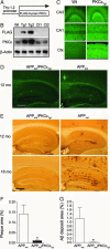
Fig. 1.
Fig. 1.
Overexpression of PKCε reduces amyloid plaque burden and inhibits Aβ accumulation in brain parenchyma. (A) Schematic of the human PKCε transgene. (B) Immunoblot of brain homogenates from wild-type mice (Wt) and from transgenic PKCε (Tg1 and Tg2) and nontransgenic (Ct1 and Ct2) littermates. (C) Similar distribution of PKCε-like immunoreactivity in hippocampal CA3 and CA1 regions and frontal cortex (Ctx) of Wt and PKCεTg1 mice. (D and E) Thioflavin-S staining (D) and anti-Aβ immunostaining (3D6 antibody) (E) showing fewer plaques and Aβ immunoreactive deposits in the hippocampus and neocortex in APPInd/PKCεTg1 mice than in APPInd mice. (Scale bars: 200 μm.) (F and G) Quantification of Thioflavin-S staining (F) and Aβ deposits (G) is expressed as the percentage of total surface area of the hippocampus and cortex in each section. Data are from 10 mice per genotype, averaged from three sections per mouse. ∗, P < 0.05 by two-tailed t tests.
Fig. 2.
Fig. 2.
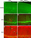
PKCε decreases plaque-associated neuropathology. (A and B) Patches of GFAP-positive astrocytes were detected by confocal microscopy in the parietal cortex in singly transgenic APPInd mice but not in doubly transgenic APPInd/PKCεTg1 mice. (C and D) Dual-channel immunofluorescence images (anti-APP, green; GFAP, red) show astrocytosis and disruption of the pyramidal cell layer in the hippocampus of an APPInd mouse and no such changes in an APPInd/PKCεTg1 mouse. (E and F) Dual-channel immunofluorescence images (anti-APP, green; MAP-2, red) demonstrating plaques (arrows) and displacement of dendrites in the CA1 region of an APPInd mouse but not in an APPInd/PKCεTg1 mouse. (Scale bars: AD, 100 μm; E and F, 50 μm.)
Fig. 3.
Fig. 3.
Overexpression of PKCε does not change levels of αAPPs and C-terminal fragments (CTFs) of APP. (A Upper) A representative immunoblot of sAPP and flAPP immunoreactivity from hippocampi of individual mice. (A Lower) The quantification of immunoreactive bands, indicating no difference between the genotypes (n = 7 for APPInd and n = 8 for APPInd/PKCεTg1) in relative sAPP levels, which were normalized to the level of flAPP. (B Upper) Representative immunoblots of CTF-α (C83) and CTF-β (C99) immunoreactivity. (B Lower) No difference in levels of these CTFs between APPInd and APPInd/PKCεTg1 mice (n = 5 for each genotype). CTF immunoreactivity was normalized to GAPDH immunoreactivity.
Fig. 4.
Fig. 4.
Effect of PKCε on Aβ-degrading enzymes. (A and B) IDE (A) and NEP (B) activity in the forebrain were similar in APPInd mice (n = 3) and APPInd/PKCεTg1 mice (n = 5). (C and D) ECE activity in the parietal cortex (C) and hippocampus (D) of APPInd mice (n = 7) and APPInd/PKCεTg1 mice (n = 8). (E) PKCε levels were determined by Western blot analysis (Left) and ECE (Right) activity in PKCε-transfected (n = 12) and mock-transfected EA.hy926 cultures (n = 12) after treatment with 16 nM phorbol 12-myristate 13-acetate for 24 h. ∗, P < 0.05 by two-tailed, unpaired t tests in CE.

Comment in

  • Another weapon against amyloid.
    Greenberg DA. Greenberg DA. Proc Natl Acad Sci U S A. 2006 May 23;103(21):7943-4. doi: 10.1073/pnas.0602860103. Epub 2006 May 16. Proc Natl Acad Sci U S A. 2006. PMID: 16705031 Free PMC article. No abstract available.

Similar articles

Cited by

References

    1. Selkoe D. J. Physiol. Rev. 2001;81:741–766. - PubMed
    1. Xu H., Greengard P., Gandy S. J. Biol. Chem. 1995;270:23243–23245. - PubMed
    1. Skovronsky D. M., Moore D. B., Milla M. E., Doms R. W., Lee V. M. J. Biol. Chem. 2000;275:2568–2575. - PubMed
    1. Savage M. J., Trusko S. P., Howland D. S., Pinsker L. R., Mistretta S., Reaume A. G., Greenberg B. D., Siman R., Scott R. W. J. Neurosci. 1998;18:1743–1752. - PMC - PubMed
    1. Selkoe D. J., Schenk D. Annu. Rev. Pharmacol. Toxicol. 2003;43:545–584. - PubMed

Publication types

LinkOut - more resources