. 2006 May 23;103(21):8227-32.
doi: 10.1073/pnas.0509400103. Epub 2006 May 12.

Synaptic tetraspan vesicle membrane proteins are conserved but not needed for synaptogenesis and neuronal function in Caenorhabditis elegans

Affiliations

Synaptic tetraspan vesicle membrane proteins are conserved but not needed for synaptogenesis and neuronal function in Caenorhabditis elegans

Christian Abraham et al. Proc Natl Acad Sci U S A. .

Abstract

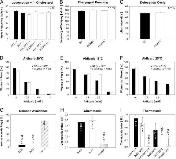
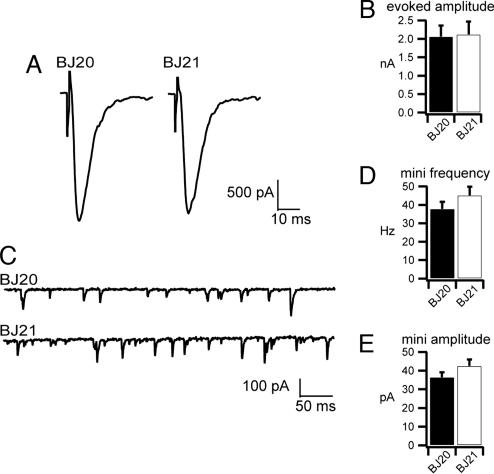
Tetraspan vesicle membrane proteins (TVPs) comprise a major portion of synaptic vesicle proteins, yet their contribution to the synaptic vesicle cycle is poorly understood. TVPs are grouped in three mammalian gene families: physins, gyrins, and secretory carrier-associated membrane proteins (SCAMPs). In Caenorhabditis elegans, only a single member of each of these families exists. These three nematode TVPs colocalize to the same vesicular compartment when expressed in mammalian cells, suggesting that they could serve overlapping functions. To examine their function, C. elegans null mutants were isolated for each gene, and a triple mutant was generated. Surprisingly, these animals develop normally and exhibit normal neuronal architecture and synaptic contacts. In addition, functions of the motor and sensory systems are normal as determined by pharmacological, chemotaxis, and thermotaxis assays. Finally, direct electrophysiological analysis of the neuromuscular junction revealed no phenotype in the TVP mutants. We therefore conclude that TVPs are not needed for the basic neuronal machinery and instead may contribute to subtle higher order functions.


Conflict of interest statement

Conflict of interest statement: No conflicts declared.

Figures

Fig. 1.
Fig. 1.
Microscopy of human epithelial PLC cells producing fluorescent C. elegans TVP fusions. Images of triple-transfected cells were recorded by epifluorescence microscopy and the insets were recorded by confocal microscopy. Color calibration bars show the degree of colocalization for each overlay.
Fig. 2.
Fig. 2.
Tissue distribution of TVPs in C. elegans. Yellow fluorescent protein (YFP) expressed under the control of the indicated promoters. An inverted fluorescence micrograph of the head of an adult is shown in each panel; anterior is up. SCAMP is expressed in an amphidial neuron (A), synaptophysin in the pharyngeal muscle cells (B), and synaptogyrin in neurons. (C). (Scale bars: 10 μm.)
Fig. 3.
Fig. 3.
Schematic representation of the TVP genes scm-1, sph-1, and sng-1 and their corresponding polypeptides, SCM-1, SPH-1, and SNG-1, together with the mutated alleles hd30 (deletion from 28,951–30,679 in AF003739), ox278 (deletion from16,583–17,122 in AF038618), ox277 (deletion from 16,805–17,151 in AF038618), and ok234 (deletion from15,736–17,437 in U40417). For each, top portion shows the protein structure, exons are colored boxes, bars denote the transmembrane domains, and dotted lines indicate deleted regions (Δ).
Fig. 4.
Fig. 4.
Neuronal architecture is normal in the TVP triple mutant. (A) Fluorescence micrographs (negative image) of the distribution of GABA neurons expressing GFP under the control of the unc-47 promoter in wild-type (wt) and mutant (mt) backgrounds (strains EG1285 and BJ1, respectively). (B) Fluorescence micrographs of SNB::GFP in the dorsal nerve cord in wild-type and mutant backgrounds (strains BJ22 and BJ28, respectively). (C) Electron microscopy of a synapse in the ventral nerve cord of a triple TVP mutant worm (strain EG2960). Note the presence of clustered synaptic vesicles close to a typical presynaptic specialization, the occurence of clathrin-coated vesicles (arrow), and the detection of an endocytotic figure (arrowhead).
Fig. 5.
Fig. 5.
Analysis of neuronal functions in wild-type (N2 and BJ20) and triple TVP mutant worms (EG2959, EG2960, and BJ21). Strains CX10 osm-9(ky10), MT3564 osm-7(n1515), and MT6308 eat-4(ky5) were used as controls in assays for osmotic avoidance, chemotaxis, and thermotaxis. Note that none of the assays revealed significant differences between wild-type and mutant animals. Scale bars depict standard error of the mean, when applicable.
Fig. 6.
Fig. 6.
Electrophysiological analysis of outcrossed triple TVP mutant worms (BJ21) and wild-type control worms (BJ20) derived from the same outcross. (A and B) Representative evoked responses (A) and mean amplitudes of evoked responses (B) (n = 5 for the wild type; n = 6 for mutant) reveal no differences (P = 0.91). (C) Representative traces of endogenous fusion events [miniature postsynaptic currents (mPSC)] in 5 mM Ca2+. (D and E) Mean frequencies of mPSCs (n = 6 each) (D), and mean amplitudes of individual miniature currents (n = 6 each) (E) were unaffected in TVP mutants (P = 0.26 and P = 0.20, respectively).

Similar articles

Cited by

References

    1. Südhof T. C. Annu. Rev. Neurosci. 2004;27:509–547. - PubMed
    1. Hübner K., Windoffer R., Hutter H., Leube R. E. Int. Rev. Cytol. 2002;214:103–159. - PubMed
    1. Eshkind L. G., Leube R. E. Cell Tissue Res. 1995;282:423–433. - PubMed
    1. McMahon H. T., Bolshakov V. Y., Janz R., Hammer R. E., Siegelbaum S. A., Südhof T. C. Proc. Natl. Acad. Sci. USA. 1996;93:4760–4764. - PMC - PubMed
    1. Fernandez-Chacon R., Alvarez de Toledo G., Hammer R. E., Südhof T. C. J. Biol. Chem. 1999;274:32551–32554. - PubMed

Publication types

LinkOut - more resources