. 2006 Jun;18(6):1412-25.
doi: 10.1105/tpc.105.039644. Epub 2006 May 12.

Gamma-tubulin is essential for microtubule organization and development in Arabidopsis

Affiliations

Gamma-tubulin is essential for microtubule organization and development in Arabidopsis

Martine Pastuglia et al. Plant Cell. 2006 Jun.

Abstract

The process of microtubule nucleation in plant cells is still a major question in plant cell biology. gamma-Tubulin is known as one of the key molecular players for microtubule nucleation in animal and fungal cells. Here, we provide genetic evidence that in Arabidopsis thaliana, gamma-tubulin is required for the formation of spindle, phragmoplast, and cortical microtubule arrays. We used a reverse genetics approach to investigate the role of the two Arabidopsis gamma-tubulin genes in plant development and in the formation of microtubule arrays. Isolation of mutants in each gene and analysis of two combinations of gamma-tubulin double mutants showed that the two genes have redundant functions. The first combination is lethal at the gametophytic stage. Disruption of both gamma-tubulin genes causes aberrant spindle and phragmoplast structures and alters nuclear division in gametophytes. The second combination of gamma-tubulin alleles affects late seedling development, ultimately leading to lethality 3 weeks after germination. This partially viable mutant combination enabled us to follow dynamically the effects of gamma-tubulin depletion on microtubule arrays in dividing cells using a green fluorescent protein marker. These results establish the central role of gamma-tubulin in the formation and organization of microtubule arrays in Arabidopsis.


Figures
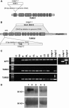
Figure 1.
Figure 1.
Isolation of T-DNA Insertions in the Two γ-Tubulin Genes and Protein Gel Blot Analysis of Double Mutant Plants. (A) and (B) Schematic representations of the mutant tubg1 and tubg2 loci indicating the position of the T-DNA insertion sites. The structure of the three loci was determined by DNA gel blot analysis, using T-DNA left and right border probes and sequencing of the T-DNA flanking regions (data not shown). A single full-length T-DNA inserted in the first exon of the TUBG1 gene is present in tubg1-1 (A). A pair of T-DNAs organized in inverted repeat is inserted in tubg2-1 (B). The T-DNA insertion induced a 2.4-kb deletion at the tubG2 loci, removing most of the coding region of TUBG2 from the end of the second exon to the end of the gene 40 bp upstream of the transcriptional start of the adjacent At5g05630 gene. At5g05630 encodes an unknown protein with weak similarity to amino acid permease family proteins. RT-PCR analysis showed that At5g05630 RNA is present in tubg2-1 plants at the same level as in wild-type plants in all tissue examined (data not shown). This result, plus the fact that tubg2-1 homozygous plants have no visible phenotype, strongly supports that the tubg2-1 insertion does not affect At5g05630 function. In the tubg2-2 line, three full-length linked T-DNAs are inserted in the TUBG2 5′ untranslated region. Gray boxes numbered I to X, exons; unnumbered gray boxes, 5′ or 3′ untranslated regions. (C) RT-PCR analysis of TUBG1 and TUBG2 transcripts in wild-type organs and in mutant plants. Primer combinations specific for TUBG1 or TUBG2 cDNAs were used for RT-PCR amplification. The constitutively expressed APRT1 gene was used as a semiquantitative control (Moffatt et al., 1994). Using a TUBG2-specific primer combination, an abundant fusion transcript is detected in the tubg2-2 mutant (bottom panel, arrow) that is also detected using a T-DNA primer and a TUBG2-specific primer (right panel, arrow). Similarly, a fusion transcript between the T-DNA and the 3′ part of the TUBG1 gene is detected in the tubg1-1 mutant. R, roots; RL, rosette leaves; S, stem; CL, cauline leaves; F, flowers; ©, suspension cultured cells; LG, light-grown whole seedlings; DG, dark-grown whole seedlings; Gn, genomic DNA. (D) Protein gel blot analysis of γ-tubulin content in tubg1-1 tubg2-2 mutant plants. Proteins were extracted from whole seedlings aged 1 week. Total protein extract (150 μg) from wild-type (left) and double mutant plants (right) was loaded on a two-well preparative SDS-PAGE gel. The gel was transferred onto a membrane, and a 16-lane multislot blot system was used for hybridization, each lane corresponding to ∼15 μg of total protein. The bottom panel shows Coomassie blue staining of the membrane, revealing the ribulose-1,5-bis-phosphate carboxylase/oxygenase (Rubisco) large subunit as a loading control. The top panel shows wild-type (lanes 1 to 3) and tubg1-1 tubg2-2 double mutant (lanes 4 and 5) extracts probed with a polyclonal anti-γ-tubulin antiserum raised against the full-length tobacco (Nicotiana tabacum) protein. The purified antibody was used at a dilution of 1:4000 (lanes 1 and 4) or 1:8000 (lanes 2, 3, and 5). Specificity was demonstrated by competition with 20 nM purified recombinant γ-tubulin (lane 3). γ-Tubulin (∼53 kD) migrates in the same region as the Rubisco large subunit, and a faint cross-hybridization of the γ-tubulin antiserum to Rubisco is visible on the protein gel blot just below the specific band, which is not competed out by addition of purified recombinant γ-tubulin (lane 3).
Figure 2.
Figure 2.
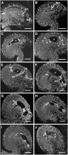
Phenotype of tubg1-1 tubg2-1 Female Gametophytes. Confocal images of wild-type control ([A] to [F]) and tubg1-1 tubg2-1 gametophytes ([G] to [J]) prepared as described by Christensen et al. (1997) (1998). The bright fluorescent spots are nucleoli and reflect the position of nuclei. All ovules have the same orientation: the chalazal pole is on the left, and the micropylar pole is on the right. ch, chalazal pole; mp, micropylar pole; dm, degenerating spores; v, central vacuole; an, antipodal nucleoli; pn, polar nucleoli; ccn, central cell nucleus; en, egg nucleolus; sn, synergids nucleoli. These images are projections of several 1-μm optical sections. Bars = 20 μm. (A) Wild-type uninucleate gametophyte: shortly after meiosis, three spores degenerate, whereas the one located at the chalazal pole expands, giving rise to the functional gametophyte. (B) The functional spore undergoes mitosis and produces a two-nucleate gametophyte visible here. Shortly afterwards, vacuoles coalesce into a large vacuole separating the two nuclei. (C) Four-nucleate gametophyte after second mitosis; the two pairs of nuclei are separated by a large central vacuole. (D) Eight-nucleate gametophyte generated by the third mitosis. The eight nuclei are indicated by arrowheads. (E) Nuclear migration in the eight-nucleate embryo sac: the two polar nuclei (previously one at the chalazal pole and one at the micropylar pole) have migrated to the micropylar half of the gametophyte where they will ultimately fuse to give the central cell nucleus visible in (F). (F) After the third mitosis, cellularization begins, giving rise to the mature female gametophyte composed of the egg cell, which will give the embryo upon fertilization, the central cell, which results from the polar nuclei fusion that will give the endosperm after fertilization, and the two synergids and three antipodal cells. The antipodal cells undergo cell death upon complete maturation. (G) to (J) Examples of abnormal gametophytes observed in mature ovules of a [TUBG1/tubg1-1; TUBG2/tubg2-1] plant (six nuclei in [G], four nuclei in [H] and [I], and three nuclei in [J]; arrowheads indicate position of antipodal nuclei). In these gametophytes, nuclei present at the central position are mostly of equal size (as judged from the size of the nucleoli), instead of a central cell nucleus and a significantly smaller egg cell nucleus in the wild type (compare with central cell nucleus and egg nucleolus in [F]).
Figure 3.
Figure 3.
Nuclear Phenotype of tubg1-1 tubg2-1 Mature Pollen. DAPI staining of mature pollen grains. The mature male gametophyte of Arabidopsis (or pollen grain) is a tricelled structure containing two sperm cells (the gametes) enclosed in a vegetative cell. During pollen development, a sporogenous cell undergoes meiosis I and II and produces a tetrad of spores. The first division of the spore is an asymmetric mitosis that gives rise to a vegetative and a sperm cell, which is then internalized. The sperm cell divides once more to produce two gametes. Upon pollination, the vegetative cell sustains growth of the pollen tube in the female tissue, enabling delivery of male gametes to the female gametophyte and double fertilization. Bars = 10 μm. (A) Typical trinucleate mature pollen grain from a wild-type hybrid control plant. The nucleus of the vegetative cell stains less densely than the two nuclei of the sperm cells that are highly fluorescent. (B) to (F) Abnormal pollen grain in an anther of a [TUBG1/tubg1-1; TUBG2/tubg2-1] plant. Most of the abnormal pollen contains only two nuclei ([B] and [C]), one appearing dispersed and similar to the vegetative one and the other one appearing densely stained. Some pollen contain two identical nuclei (E) and some only one nucleus (F).
Figure 4.
Figure 4.
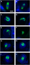
Microtubule Organization during tubg1-1 tubg2-1 Pollen Development. Overlay images of anti-α-tubulin immunolocalization (green) and DAPI staining (blue). (A) to (F) Spindle organization during the first mitosis of pollen development in anthers of wild-type hybrids ([A] and [B]) and [TUBG1/tubg1-1; TUBG2/tubg2-1] F1 plants ([C] to [F]). Metaphase (A) and anaphase (B) spindle in wild-type pollen. Spindle morphology is greatly affected in tubg1-1 tubg2-1 mutant pollen, and spindles appear as bent ([C] to [F]) or even collapsed (F) structures. (G) and (H) Abnormal microtubular structures in early two-nucleate pollen in [TUBG1/tubg1-1; TUBG2/tubg2-1] anthers. DAPI staining and nuclear morphology indicate that nuclear division has been achieved and thus strongly suggest that collapsed microtubular structures correspond to failed phragmoplasts. (I) and (J) Loss of asymmetric division during mitosis I in pollen of [TUBG1/tubg1-1; TUBG2/tubg2-1] plants (J) compared with a wild-type plant (I). During mitosis I, phragmoplast is shifted to the side of the pollen in the wild-type (I), whereas in a tubg1-1 tubg2-1 pollen, the phragmoplast is in the center of the cell. Images are stack of 1-μm optical sections. Bar = 8 μm.
Figure 5.
Figure 5.
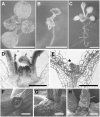
Phenotype of the Aerial Parts of Wild-Type and tubg1-1 tubg2-2 Double Mutant Seedlings. (A) and (B) Phenotype of 2-week-old wild-type (A) and tubg1-1 tubg2-2 double mutant (B) plants. Mutant plants are similar to the wild type for the first 3 to 4 days following germination and subsequently display several morphological defects, including reduced expansion and deformation of the cotyledons and defective leaf formation. (C) The phenotype of a tubg1-1 tubg2-2 double mutant is fully rescued by a 35S-TUBG2 construct. Shown here is a 3-week-old plant homozygous for the tubg1-1 and tubg2-2 mutations and heterozygous for the complementing construct. (D) and (E) Toluidine blue–stained longitudinal section in the shoot apical meristem of 10-d-old wild-type (D) and tubg1-1 tubg2-2 double mutant (E) plants. In the wild type, the shoot meristem displays small, densely stained cells (D). The corresponding region in mutant plants contains a reduced number of larger and less densely stained cells, indicating that meristem cells initiated differentiation (E). Similarly, cells in the leaf primordia (asterisk) undergo differentiation, and leaf formation is inhibited. (F) to (H) Scanning electron micrographs of the shoot apical meristem region. (F) Emergence of leaves at the shoot apex of a 7-d-old wild-type seedling. (G) At the same stage, no leaves are initiated in the tubg1-1 tubg2-2 double mutant. (H) Abnormal leaves are eventually initiated in the tubg1-1 tubg2-2 double mutant but undergo little subsequent growth, as shown in this micrograph of the meristem of an 11-d-old double mutant seedling. Bars = 5 mm in (A) to (C) and 200 μm in (D) to (H).
Figure 6.
Figure 6.
Root Growth and Morphology Defects in the tubg1-1 tubg2-2 Double Mutant. Comparison of confocal optical median sections of roots of 4-d-old wild-type (A) and mutant (B) plants and 7-d-old mutant (C) plants. In the mutant, cell expansion occurs in the root meristem (arrowhead in [B]), indicating premature arrest of cell division. The root apex thereafter undergoes radial swelling (C), as also shown by scanning electron micrographs of wild-type (D) and double mutant (E) 11-d-old seedlings. Roots were stained with FM1-43 in (A) to (C). Bars = 50 μm in (A) to (C) and 200 μm in (D) and (E).
Figure 7.
Figure 7.
Microtubule Organization in Wild-Type and tubg1-1 tubg2-2 Plants. (A) to (D) Mitotic microtubule arrays in root meristem cells of wild-type plants 4 to 7 d after germination. Microtubules are first organized in a cortical preprophase band encircling the nucleus (arrowheads in [A]). After nuclear envelope breakdown, the mitotic spindle forms (B). At the end of anaphase, the phragmoplast, constituted of two sets of opposing microtubules, appears between the spindle poles (C), expands as a ring (D), and eventually reaches the edge of the cells. (E) to (H) Microtubules arrays in root meristem cells of 4- to 7-d-old tubg1-1 tubg2-2 seedlings. Fewer mitotic microtubule arrays were observed at these stages in the mutant root meristem. Observed arrays include highly distorted or bent spindles ([E] and [F]), abnormal asymmetric phragmoplast (G), or static condensed stacks of microtubules (H). (I) to (L) Progressive disorganization of microtubule arrays after 3 to 4 d of postembryonic growth in tubg1-1 tubg2-2 mutants. Three days after germination, cortical microtubules in elongating root cells are organized normally in parallel arrays, perpendicular to the cell main axis (I). Thereafter, in the root elongation zone and early differentiation zone of 4- to 10-d-old mutant seedlings, progressive disruption of cortical microtubule arrays occurs, accompanied by radial swelling ([J] to [L]). Microtubule defects are first characterized by a loss of transverse alignment ([J] and [K]). Subsequently, cortical microtubules are depolymerized (L), as indicated by the presence of short microtubules (arrowhead) or patches of GFP fluorescence (inset) and eventually by a diffuse cytoplasmic GFP staining. Microtubule arrays are labeled by expression of the GFP-MBD fusion protein and imaged by confocal microscopy. (A) to (H) are single optical sections; (I) to (L) are stacks of multiple images taken 1 μm ([I], [J], and [L]) or 2 μm (K) apart. Bars = 10 μm in (A) to (H) and 50 μm in (I) to (L).
Figure 8.
Figure 8.
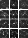
Time-Lapse Observation of a Dividing Cell in the tubg1-1 tubg2-2 Double Mutant Root Tip. (A) Preprophase band of normal appearance. (B) to (E) Abnormal mitotic spindle. (F) to (H) Abnormal asymmetric phragmoplast with two sets of opposing microtubules of unequal width. The late disorganized phragmoplast is finally rejected to the cell periphery (H). (I) and (J) Exit of mitosis without karyokinesis (only one nucleus in [I]) and without cytokinesis (absence of new membrane and cell wall deposition in [J]). The duration of the cell cycle (at least 144 min) is longer than in wild-type root tip cells (<90 min in Azimzadeh et al., 2001). This is mostly due to the persistence of the abnormal spindle (80 min from [B] to [E]) compared with duration of a typical spindle phase in the wild type (20 min in Azimzadeh et al., 2001). Microtubule arrays are revealed by the GFP-MBD fusion protein and imaged by confocal microscopy. All images are stacks of multiple images taken 0.6 μm apart. Bar = 10 μm.

Similar articles

Cited by

References

    1. Alexander, M.P. (1969). Differential staining of aborted and non aborted pollen. Stain Technol. 44 117–122. - PubMed
    1. Alonso, J.M., et al. (2003). Genome-wide insertional mutagenesis of Arabidopsis thaliana. Science 301 653–657. - PubMed
    1. Arabidopsis Genome Initiative (2000). Analysis of the genome sequence of the flowering plant Arabidopsis thaliana. Nature 408 796–815. - PubMed
    1. Azimzadeh, J., Traas, J., and Pastuglia, M. (2001). Molecular aspects of microtubule dynamics in plants. Curr. Opin. Plant Biol. 4 513–519. - PubMed
    1. Baluska, F., Hlavacka, A., Samaj, J., Palme, K., Robinson, D.G., Matoh, T., McCurdy, D.W., Menzel, D., and Volkmann, D. (2002). F-actin-dependent endocytosis of cell wall pectins in meristematic root cells. Insights from brefeldin A-induced compartments. Plant Physiol. 130 422–431. - PMC - PubMed

Publication types

LinkOut - more resources