. 2006 Jun;80(11):5125-34.
doi: 10.1128/JVI.02674-05.

Epstein-Barr virus protein kinase BGLF4 is a virion tegument protein that dissociates from virions in a phosphorylation-dependent process and phosphorylates the viral immediate-early protein BZLF1

Affiliations

Epstein-Barr virus protein kinase BGLF4 is a virion tegument protein that dissociates from virions in a phosphorylation-dependent process and phosphorylates the viral immediate-early protein BZLF1

Risa Asai et al. J Virol. 2006 Jun.

Abstract

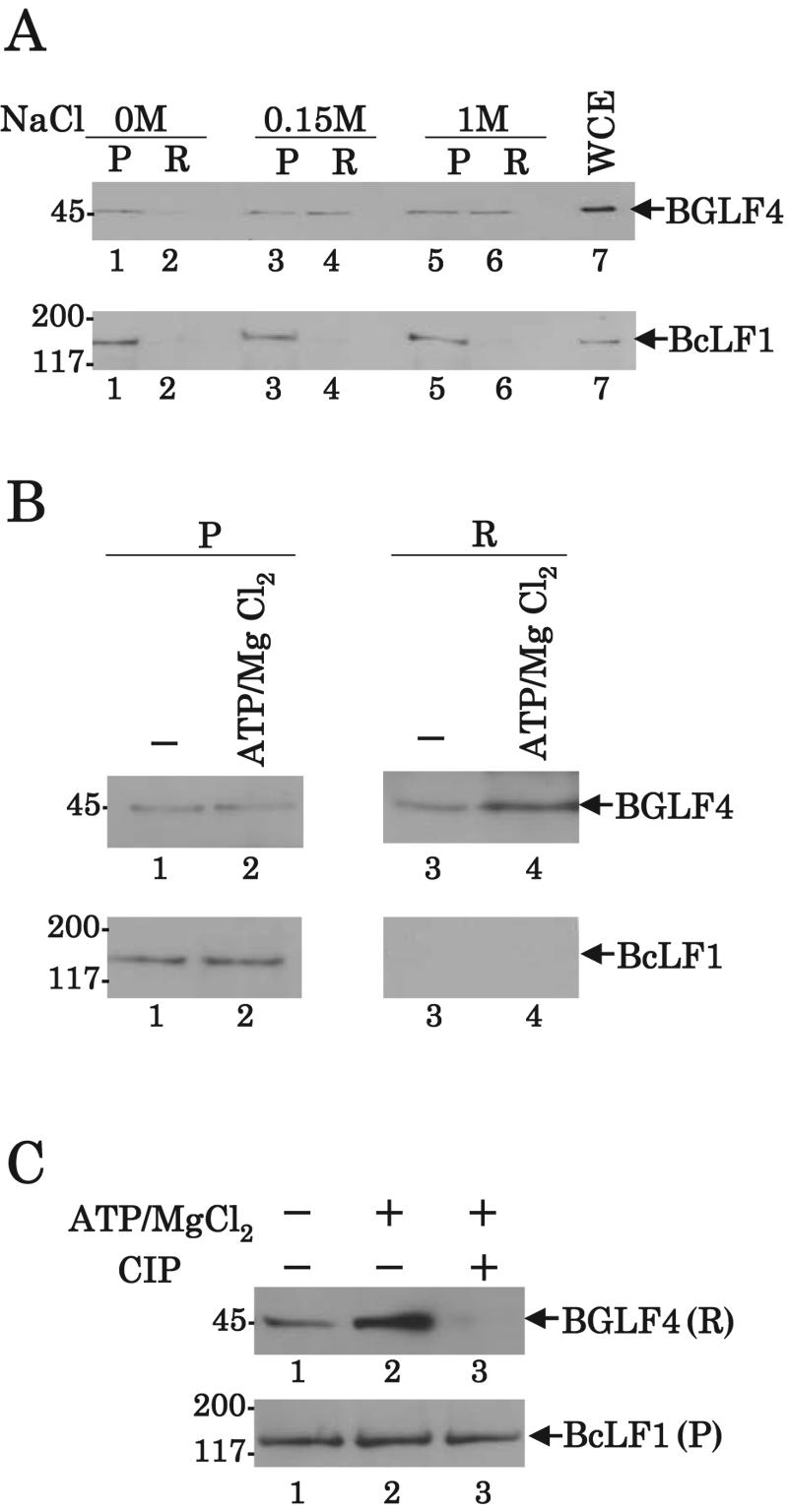
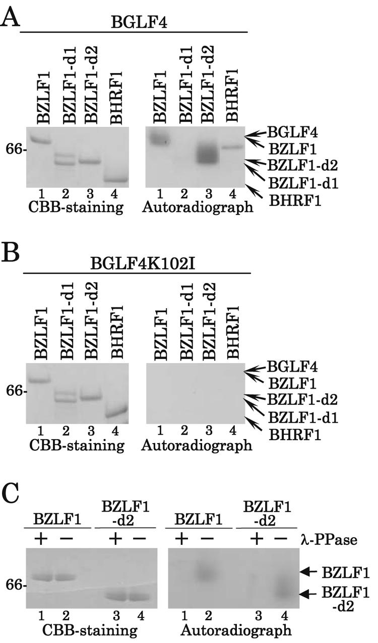
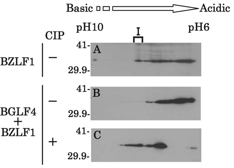
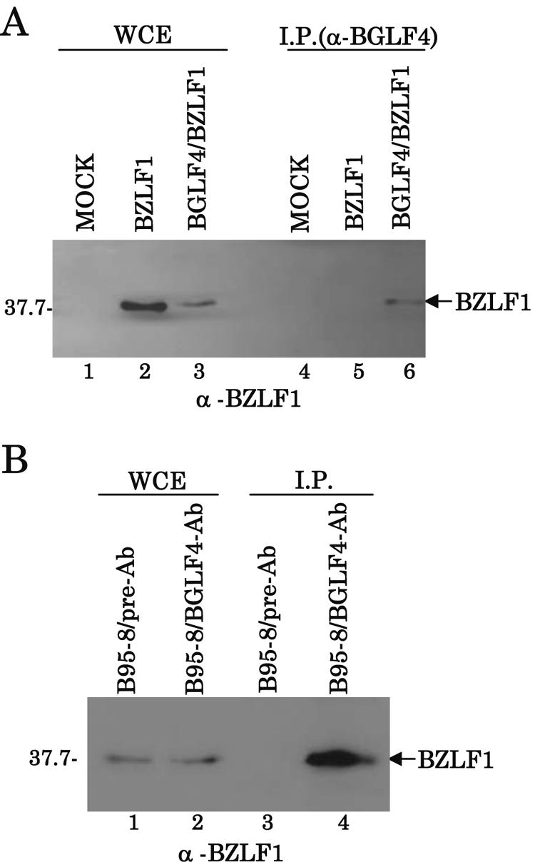
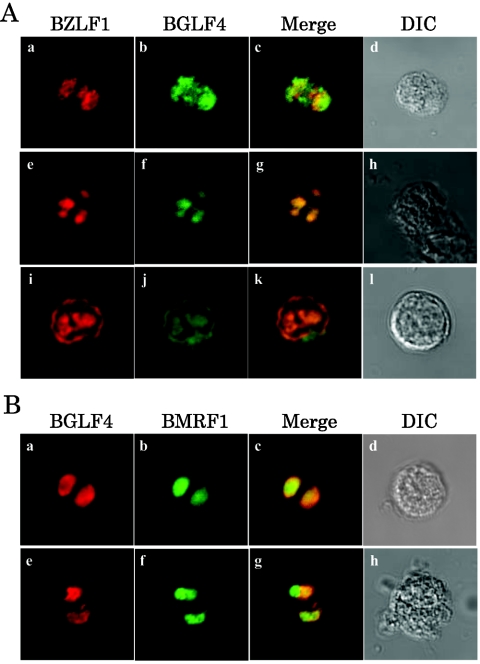
Epstein-Barr virus (EBV) BGLF4 is a viral protein kinase that is expressed in the lytic phase of infection and is packaged in virions. We report here that BGLF4 is a tegument protein that dissociates from the virion in a phosphorylation-dependent process. We also present evidence that BGLF4 interacts with and phosphorylates BZLF1, a key viral regulator of lytic infection. These conclusions are based on the following observations. (i) In in vitro tegument release assays, a significant fraction of BGLF4 was released from virions in the presence of physiological NaCl concentrations. (ii) Addition of physiological concentrations of ATP and MgCl(2) to virions enhanced BGLF4 release, but phosphatase treatment of virions significantly reduced BGLF4 release. (iii) A recombinant protein containing a domain of BZLF1 was specifically phosphorylated by purified recombinant BGLF4 in vitro, and BGLF4 altered BZLF1 posttranslational modification in vivo. (iv) BZLF1 was specifically coimmunoprecipitated with BGLF4 in 12-O-tetradecanoylphorbol-13-acetate-treated B95-8 cells and in COS-1 cells transiently expressing both of these viral proteins. (v) BGLF4 and BZLF1 were colocalized in intranuclear globular structures, resembling the viral replication compartment, in Akata cells treated with anti-human immunoglobulin G. Our results suggest that BGLF4 functions not only in lytically infected cells by phosphorylating viral and cellular targets but also immediately after viral penetration like other herpesvirus tegument proteins.


Figures

FIG. 1.
FIG. 1.
Schematic diagram of the EBV genome and location of the genes studied in this report. Line 1: diagram of the EBV genome. The unique sequences are designated U1 to U5. The terminal and internal repeats flanking the unique sequences are shown as open rectangles with their designations above the rectangles. Line 2: expanded section of the domains encoding BGLF4 and BcLF1. The polarity and structure of the BGLF4 and BcLF1 coding regions are shown. Line 3: diagram of the 1,547 codons of BcLF1. Line 4: diagram of the BcLF1 peptides used to generate GST-BcLF1 fusion proteins. Line 5: expanded diagram of the domain encoding BZLF1. Line 6: diagram of the 245 codons of BZLF1. Line 7: diagram of the BZLF1 peptides used to generate MBP-BZLF1 fusion proteins.
FIG. 2.
FIG. 2.
Analysis of virion-associated BGLF4. (A and B) Immunoblots of electrophoretically separated lysates from Ramos (lane 1) and B95-8 cells without (lane 2) or with (lane 3) TPA treatment. Cell lysates were analyzed by immunoblotting with polyclonal antibody to BGLF4 (A) and BcLF1 (B). (C and D) Immunoblots of electrophoretically separated sucrose gradient fractions of BGLF4 (C) and BcLF1 (D). As described in Materials and Methods, virions harvested from the culture supernatants of TPA-treated B95-8 cells were separated in a discontinuous sucrose gradient. Fractions were collected, separated by electrophoresis, and immunoblotted with polyclonal antibody to BGLF4 (C) and BcLF1 (D). Lanes 1 and 2, whole-cell extracts of TPA-treated B95-8 and Ramos cells, respectively. Numbers at left are molecular masses in kilodaltons.
FIG. 3.
FIG. 3.
Autoradiographs of BGLF4 immunoprecipitates after in vitro kinase assays and electrophoresis. (A) Purified virions from one of the virion-containing fractions described in Fig. 2C and D (lane 1) and from a corresponding fraction from the supernatant of Ramos cells (lane 2) were lysed and immunoprecipitated with antibody to BGLF4. The immunoprecipitates were incubated in kinase buffer containing [γ-32P]ATP, separated on a denaturing gel, transferred to a nitrocellulose membrane, and analyzed by autoradiography. (B) Immunoblot of the nitrocellulose membrane in panel A using anti-BGLF4 antibody. (C) Immunoprecipitates prepared as in panel A either mock treated (lane 1) or treated with λ-PPase (lane 2), separated on a denaturing gel, transferred to a nitrocellulose membrane, and analyzed by autoradiography. (D) Immunoblot of the nitrocellulose membrane in panel C using anti-BGLF4 antibody. Numbers at left are molecular masses in kilodaltons.
FIG. 4.
FIG. 4.
Immunoblots of in vitro tegument release assays of BGLF4 and BcLF1. (A) EBV virions from the supernatant of TPA-treated B95-8 cells were treated with Triton X-100 in 0, 0.15, or 1 M NaCl. The samples were then separated by sucrose gradient centrifugation into pelleted (P) and released (R) fractions and analyzed by immunoblotting with antibody to BGLF4 (upper panel) or BcLF1 (lower panel). Lane 7 contains whole-cell extract (WCE). (B) EBV virions were treated with lysis buffer B in the absence or presence of 1 mM ATP and 1 mM MgCl2 at 37°C for 1 h, fractionated as described in panel A, and analyzed by immunoblotting with antibody to BGLF4 (upper panels) or BcLF1 (lower panels). (C) EBV virions were treated with lysis buffer B with or without ATP and MgCl2 in the absence or presence of CIP at 37°C for 1 h, fractionated as described in panel A, and analyzed by immunoblotting with antibody to BGLF4 or BcLF1. The fractions of released (R) BGLF4 (upper panel) and pelleted (P) BcLF1 (lower panel) are shown. Numbers at left are molecular masses in kilodaltons.
FIG. 5.
FIG. 5.
Autoradiographs of in vitro BZLF1 phosphorylation. (A and B) Left panels: CBB-stained gels of phosphorylated BZLF1. Purified MBP-BZLF1 (lane 1), MBP-BZLF1d1 (lane 2), MBP-BZLF1d2 (lane 3), and MBP-BHRF1 (lane 4) were incubated in kinase buffer containing [γ-32P]ATP and purified GST-BGLF4 (A) or GST-BGLF4K102I (B), separated on a denaturing gel, and stained with CBB. Right panels: autoradiographs of the gels in the left panels. (C) Left panel: purified MBP-BZLF1 (lanes 1 and 2) and MBP-BZLF1d2 (lanes 3 and 4) incubated in kinase buffer containing [γ-32P]ATP and purified GST-BGLF4 were either mock treated (lanes 2 and 4) or treated with λ-PPase (lanes 1 and 3), separated on a denaturing gel, and stained with CBB. Right panel: autoradiograph of the gel in the left panel. Numbers at left are molecular masses in kilodaltons.
FIG. 6.
FIG. 6.
Effect of BGLF4 expression on posttranslational modification of BZLF1 protein in COS-1 cells. COS-1 cells transiently expressing either BZLF1 alone (A) or both BZLF1 and BGLF4 (B and C) were solubilized, mock treated (A and B) or treated with CIP (C), separated by two-dimensional electrophoresis, and immunoblotted with mouse monoclonal antibody to BZLF1. Numbers at left are molecular masses in kilodaltons.
FIG. 7.
FIG. 7.
Interaction of BGLF4 with BZLF1. (A) COS-1 cells mock transfected (lane 4) or transiently expressing BZLF1 alone (lane 5) or both BGLF4 and BZLF1 (lane 6) were solubilized and immunoprecipitated with antibody to BGLF4. The immunoprecipitates were analyzed by electrophoresis and immunoblotted with antibody to BZLF1. One-sixtieth of the COS-1 whole-cell extract (WCE) used in the reaction mixtures for lanes 4, 5, and 6 was loaded in lanes 1, 2, and 3, respectively. (B) TPA-treated B95-8 cells were solubilized and immunoprecipitated with anti-BGLF4 antibody (lane 4) or preimmune serum (lane 3). The immunoprecipitates were analyzed by electrophoresis and immunoblotted with mouse monoclonal antibody to BZLF1. One-sixtieth of the B95-8 whole-cell extract (WCE) used in the reaction mixtures for lanes 3 and 4 was loaded in lanes 1 and 2, respectively. Numbers at left are molecular masses in kilodaltons.
FIG. 8.
FIG. 8.
Immunofluorescent localization of BGLF4, BZLF1, and BMRF1 in EBV-infected cells. (A) Digital confocal microscope images showing localization of BGLF4 and BZLF1 in Akata cells treated with anti-human IgG for 9 h. The cells were then fixed, permeabilized, and double labeled with a combination of mouse monoclonal antibody to BZLF1 (a, e, and i) and rabbit polyclonal antibody to BGLF4 (b, f, and j), which were detected with Alexa 546-conjugated anti-mouse IgG antibody (red fluorescence) and FITC-conjugated anti-rabbit IgG antibody (green fluorescence), respectively. Single-color images were captured separately and are shown in panels a, b, e, f, i, and j. Panels c, g, and k and panels d, h, and l show simultaneous acquisitions of both colors and differential interference contrast (DIC), respectively. (B) Digital confocal microscope images showing localization of BGLF4 and BMRF1 in Akata cells treated with anti-human IgG for 9 h. The cells were then processed as described in panel A and double labeled with a combination of rabbit polyclonal antibody to BGLF4 (a and e) and mouse monoclonal antibody to BMRF1 (b and f), which were detected with Alexa 546-conjugated anti-mouse IgG antibody (red fluorescence) and FITC-conjugated anti-rabbit IgG antibody (green fluorescence), respectively. Single-color images were captured separately and are shown in panels a, b, e, and f. Panels c and g and panels d and h show simultaneous acquisitions of both colors and differential interference contrast, respectively.

References

    1. Baumann, M., H. Mischak, S. Dammeier, W. Kolch, O. Gires, D. Pich, R. Zeidler, H. J. Delecluse, and W. Hammerschmidt. 1998. Activation of the Epstein-Barr virus transcription factor BZLF1 by 12-O-tetradecanoylphorbol-13-acetate-induced phosphorylation. J. Virol. 72:8105-8114. - PMC - PubMed
    1. Chee, M. S., G. L. Lawrence, and B. G. Barrell. 1989. Alpha-, beta- and gammaherpesviruses encode a putative phosphotransferase. J. Gen. Virol. 70:1151-1160. - PubMed
    1. Chen, M. R., S. J. Chang, H. Huang, and J. Y. Chen. 2000. A protein kinase activity associated with Epstein-Barr virus BGLF4 phosphorylates the viral early antigen EA-D in vitro. J. Virol. 74:3093-3104. - PMC - PubMed
    1. Cohen, J. I., and S. E. Straus. 2001. Varicella-zoster virus and its replication, p. 2707-2730. In D. M. Knipe, P. M. Howley, D. E. Griffin, R. A. Lamb, M. A. Martin, B. Roizman, and S. E. Straus (ed.), Fields virology, 4th ed. Lippincott-Williams & Wilkins, Philadelphia, Pa.
    1. Edelman, A. M., D. K. Blumenthal, and E. G. Krebs. 1987. Protein serine/threonine kinases. Annu. Rev. Biochem. 56:567-613. - PubMed

Publication types

MeSH terms

Substances

LinkOut - more resources