. 2006 Jun;80(11):5156-67.
doi: 10.1128/JVI.02349-05.

Ebola virus VP24 binds karyopherin alpha1 and blocks STAT1 nuclear accumulation

Affiliations

Ebola virus VP24 binds karyopherin alpha1 and blocks STAT1 nuclear accumulation

St Patrick Reid et al. J Virol. 2006 Jun.

Abstract

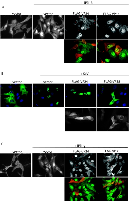
Ebola virus (EBOV) infection blocks cellular production of alpha/beta interferon (IFN-alpha/beta) and the ability of cells to respond to IFN-alpha/beta or IFN-gamma. The EBOV VP35 protein has previously been identified as an EBOV-encoded inhibitor of IFN-alpha/beta production. However, the mechanism by which EBOV infection inhibits responses to IFNs has not previously been defined. Here we demonstrate that the EBOV VP24 protein functions as an inhibitor of IFN-alpha/beta and IFN-gamma signaling. Expression of VP24 results in an inhibition of IFN-induced gene expression and an inability of IFNs to induce an antiviral state. The VP24-mediated inhibition of cellular responses to IFNs correlates with the impaired nuclear accumulation of tyrosine-phosphorylated STAT1 (PY-STAT1), a key step in both IFN-alpha/beta and IFN-gamma signaling. Consistent with this proposed function for VP24, infection of cells with EBOV also confers a block to the IFN-induced nuclear accumulation of PY-STAT1. Further, VP24 is found to specifically interact with karyopherin alpha1, the nuclear localization signal receptor for PY-STAT1, but not with karyopherin alpha2, alpha3, or alpha4. Overexpression of VP24 results in a loss of karyopherin alpha1-PY-STAT1 interaction, indicating that the VP24-karyopherin alpha1 interaction contributes to the block to IFN signaling. These data suggest that VP24 is likely to be an important virulence determinant that allows EBOV to evade the antiviral effects of IFNs.


Figures

FIG. 1.
FIG. 1.
Ebola virus VP24 inhibits IFN-β and -γ induced gene expression. (A) IFN-β-induced reporter gene activation in Vero cells was measured. Cells were cotransfected with the ISG54-CAT reporter plasmid, a constitutively expressed Renilla luciferase reporter plasmid and empty vector (Vec) or with plasmids expressing Nipah virus W protein (W), Flag-tagged EBOV VP24 (F-24), untagged VP24 (24), or EBOV VP35 (35). Twenty-four hours posttransfection, the cells either were mock treated or were treated with 1,000 U of human IFN-β/ml for 24 h, harvested, and then assayed for CAT and luciferase activities. The data are presented as the activation (fold) relative to empty vector, mock-treated controls. Error bars indicate means ± the standard deviation of three experiments. (B) Levels of STAT1 gene expression in 293T cells transfected with empty vector (Vec) or plasmids expressing the indicated protein are shown. Twenty-four hours posttransfection, the cells were treated with 1,000 U of IFN-β/ml, and 18 h posttreatment, the cells were lysed and subjected to Western blot analysis with anti-STAT1 (αSTAT1) and anti-β-actin (α β-actin) antibodies. (C) Levels of IFN-γ-induced reporter gene activation in Vero cells transfected with empty vector (Vec) or with plasmids expressing EBOV VP35 (35) or FLAG-tagged EBOV VP24 (24). Vero cells were transfected with the IFN-γ-responsive IRF-1-promoter luciferase reporter and a constitutively expressed Renilla luciferase reporter. Twenty-four hours posttransfection, the cells were either mock treated or treated with 1,000 U/ml of IFN-γ, and 24 h posttreatment, reporter gene activity was measured. IFN-γ-induced reporter values were normalized to the Renilla luciferase reporter. Results are presented as induction (fold) of the IRF-1-luciferase reporter relative to an empty vector-transfected, mock-treated control. Error bars indicate the mean ± the standard deviation of three experiments.
FIG. 2.
FIG. 2.
Ebola virus VP24 rescues growth of NDV-GFP in cells pretreated with IFN-β. Vero cells were transfected with the empty vector (Vector) or plasmids expressing the Nipah virus W (Nipah W) or EBOV VP24 (VP24) proteins. Twenty-four hours posttransfection, the cells were mock treated (−IFNβ) or treated with 1,000 U/ml of IFN-β (+IFNβ) as indicated. Twenty-four hours post-IFN-β treatment, the cells were infected with NDV-GFP. The green fluorescence was visualized 24 h postinfection under a fluorescence microscope.
FIG. 3.
FIG. 3.
EBOV protein VP24 prevents IFN-mediated nuclear translocation of STAT1. (A) IFN-β treatment (30 min) of Vero cells causes STAT1 to relocate from the cytoplasm (vector) to the nucleus (vector + IFN-β). In cells expressing FLAG-VP24 (FLAG-VP24 + IFN-β; relevant cells marked with an asterisk), STAT1 fails to relocate to the nucleus after IFN-β treatment. IFN-β treatment of FLAG-VP35-expressing cells (FLAG-VP35 + IFN-β; relevant cells marked with an asterisk) causes STAT1 to relocate to the nucleus. Upper panels show only STAT1 images. Lower panels show the STAT1 images (green) merged with images of FLAG-tagged Ebola virus proteins (red). (B) Vero cells express GFP-IRF-3 in the cytoplasm in the absence of viral infection (vector), but translocate GFP-IRF-3 to the nucleus when infected with Sendai virus (vector + SeV). Coexpression of FLAG-VP35 prevents translocation of GFP-IRF-3 (FLAG-VP35 + SeV), but FLAG-VP24-expressing cells are still able to traffic GFP-IRF-3 to the nucleus (FLAG-VP24 + SeV). Upper panels show the GFP-IRF-3 (green) merged with Hoechst nuclear staining (blue). Lower panels show the FLAG-tagged Ebola virus proteins. (C) In Vero cells cotransfected with empty vector, STAT1 is predominately cytoplasmic in the absence of IFN-γ treatment (vector), but STAT1 concentrates in the nucleus after a 30-min treatment with IFN-γ (vector + IFN-γ). In the presence of FLAG-VP24, IFN-γ treatment fails to relocate STAT1 to the nucleus (FLAG-VP24 + IFN-γ).
FIG. 4.
FIG. 4.
VP24 prevents the nuclear translocation of phosphorylated STAT1 (Tyr 701). (A) After treatment with IFN-β for 30 min, cells expressing FLAG-VP35 (top row, red channel) are able to translocate PY-STAT1 (P-STAT1; green channel) to the nucleus. In cells expressing FLAG-VP24 (bottom row, red channel) PY-STAT1 is located in the cytoplasm. (B) VP24 expression does not detectably inhibit the tyrosine phosphorylation of STAT1. 293T cells were transfected with expression plasmids encoding: pCAGGS empty vector (vector), luciferase, Nipah virus V (NipV), VP35, VP24, or FLAG-VP24. After treatment with IFN-β (or mock treatment), the cells were lysed and analyzed for the presence of PY-STAT1 by Western blotting.
FIG. 5.
FIG. 5.
EBOV infection inhibits IFN-β-induced expression of MxA and prevents nuclear translocation of PY-STAT1. (A) Mock-infected Vero cells express MxA in response to overnight treatment with IFN-β (lane 2). However, EBOV-infected cells (MOI = 1) do not produce MxA in response to IFN-β (lane 4). Lanes 1 and 2 were mock infected. Lanes 3 and 4 were infected with EBOV. Lanes 2 and 4 were treated with IFN-β, while lanes 1 and 3 were mock treated. (Top panel) Western blot to detect MxA and GAPDH. (Bottom panel) Western blot to detect VP24. (B) Mock-infected Vero cells translocate PY-STAT1 to the nucleus after 30 min of IFN-β treatment (top panel), but EBOV-infected cells retain PY-STAT1 in the cytoplasm (bottom panel). Red represents viral antigen (VP35), and green represents PY-STAT1.
FIG. 6.
FIG. 6.
VP24 interacts with karyopherin α1 and inhibits karopherin α1-phospho (Tyr 701)-STAT1 interaction. (A) Coimmunoprecipitation of VP24 with karyopherin α1. 293T cells were transfected with the indicated plasmids expressing VP24, VP35, or firefly luciferase (indicated by a hyphen) in the absence or presence of plasmids expressing FLAG-tagged karyopherin α1 (FLAG-Kα1), α2 (FLAG-Kα2), α3 (FLAG-Kα3), or α4 (FLAG-Kα4). Cell extracts were prepared 1 day posttransfection, and immunoprecipitations (IP) were performed with anti-FLAG monoclonal antibody M2 bound to agarose beads. After washing, immunoprecipitates were analyzed by Western blotting (WB) with polyclonal rabbit antisera recognizing FLAG and VP24 (top panel; 50% of total material was analyzed). Whole-cell extracts (WCE) (1% of total) were also analyzed by Western blotting for karyopherin expression (anti-FLAG antibody), VP24, or VP35, as indicated. (B) 293T cells were transfected with the indicated expression plasmids. Lanes 1 and 4, FLAG-karyopherin α1 and STAT1-GFP; lanes 2 and 5, FLAG-karyopherin α1 and HA-VP24; lanes 3 and 6, FLAG-karyopherin α1, STAT1-GFP, and HA-VP24. The cells were then mock treated or treated with 1,000 U/ml human IFN-β and subsequently immunoprecipitated with anti-FLAG monoclonal antibody M2 bound to agarose beads (IP: FLAG). After washing, the immunoprecipitates were analyzed by Western blotting with a monoclonal antibody recognizing STAT1 (WB: STAT1), and monoclonal antibodies against FLAG (WB: FLAG) and HA (WB: HA) (top panel; 10% of total material analyzed). Whole-cell extracts (1% of total) were also analyzed similarly. (C) 293T cells were transfected with FLAG-karyopherin α1 (250 ng); STAT1-GFP (250 ng); or 0, 25, 250, 500, and 1,000 ng of HA-VP24 (lanes 1 to 5, respectively) expression plasmids. Lane 6 contains samples derived from cells transfected with STAT1-GFP plasmid but not with karyopherin α1 plasmid. The cells were then treated with 1,000 U/ml human IFN-β and subsequently immunoprecipitated with anti-FLAG monoclonal antibody and analyzed by Western blotting with polyclonal rabbit antisera recognizing phosphorylated STAT1 and monoclonal antibodies against FLAG and HA (top panel, 10% of total material analyzed). Whole-cell extracts (1% of total) were also analyzed. Asterisks indicate the heavy chain of the anti-FLAG monoclonal M2 antibody used for the immunoprecipitation.

References

    1. Aebi, M., J. Fäh, N. Hurt, C. E. Samuel, D. Thomis, L. Bazzigher, J. Pavlovic, O. Haller, and P. Staeheli. 1989. cDNA structures and regulation of two interferon-induced human Mx proteins. Mol. Cell. Biol. 9:5062-5072. - PMC - PubMed
    1. Baize, S., E. M. Leroy, E. Mavoungou, and S. P. Fisher-Hoch. 2000. Apoptosis in fatal Ebola infection. Does the virus toll the bell for immune system? Apoptosis 5:5-7. - PubMed
    1. Bamberg, S., L. Kolesnikova, P. Möller, H.-D. Klenk, and S. Becker. 2005. VP24 of Marburg virus influences formation of infectious particles. J. Virol. 79:13421-13433. - PMC - PubMed
    1. Reference deleted.
    1. Basler, C. F., A. Mikulasova, L. Martinez-Sobrido, J. Paragas, E. Mühlberger, M. Bray, H.-D. Klenk, P. Palese, and A. Garcia-Sastre. 2003. The Ebola virus VP35 protein inhibits activation of interferon regulatory factor 3. J. Virol. 77:7945-7956. - PMC - PubMed

Publication types