. 2006 Jun;80(11):5168-78.
doi: 10.1128/JVI.02199-05.

Ebola virus VP35 protein binds double-stranded RNA and inhibits alpha/beta interferon production induced by RIG-I signaling

Affiliations

Ebola virus VP35 protein binds double-stranded RNA and inhibits alpha/beta interferon production induced by RIG-I signaling

Washington B Cárdenas et al. J Virol. 2006 Jun.

Abstract

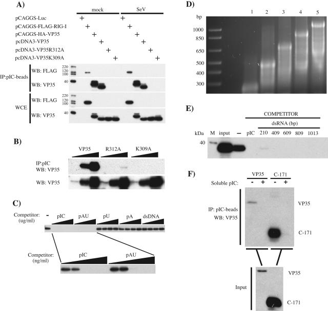
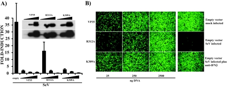
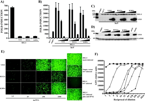
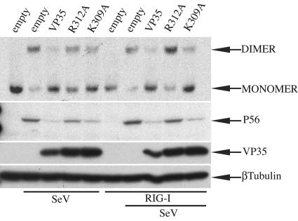
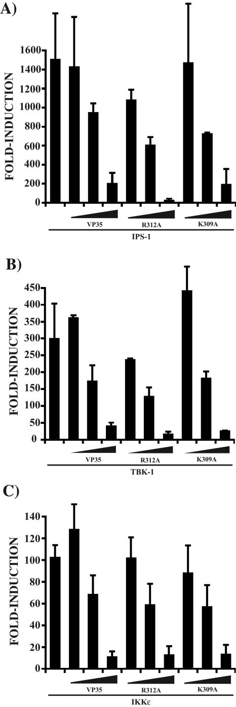
The Ebola virus (EBOV) VP35 protein blocks the virus-induced phosphorylation and activation of interferon regulatory factor 3 (IRF-3), a transcription factor critical for the induction of alpha/beta interferon (IFN-alpha/beta) expression. However, the mechanism(s) by which this blockage occurs remains incompletely defined. We now provide evidence that VP35 possesses double-stranded RNA (dsRNA)-binding activity. Specifically, VP35 bound to poly(rI) . poly(rC)-coated Sepharose beads but not control beads. In contrast, two VP35 point mutants, R312A and K309A, were found to be greatly impaired in their dsRNA-binding activity. Competition assays showed that VP35 interacted specifically with poly(rI) . poly(rC), poly(rA) . poly(rU), or in vitro-transcribed dsRNAs derived from EBOV sequences, and not with single-stranded RNAs (ssRNAs) or double-stranded DNA. We then screened wild-type and mutant VP35s for their ability to target different components of the signaling pathways that activate IRF-3. These experiments indicate that VP35 blocks activation of IRF-3 induced by overexpression of RIG-I, a cellular helicase recently implicated in the activation of IRF-3 by either virus or dsRNA. Interestingly, the VP35 mutants impaired for dsRNA binding have a decreased but measurable IFN antagonist activity in these assays. Additionally, wild-type and dsRNA-binding-mutant VP35s were found to have equivalent abilities to inhibit activation of the IFN-beta promoter induced by overexpression of IPS-1, a recently identified signaling molecule downstream of RIG-I, or by overexpression of the IRF-3 kinases IKKepsilon and TBK-1. These data support the hypothesis that dsRNA binding may contribute to VP35 IFN antagonist function. However, additional mechanisms of inhibition, at a point proximal to the IRF-3 kinases, most likely also exist.


Figures

FIG. 1.
FIG. 1.
Ebola virus VP35 binds to dsRNA. (A) 293T cells were transfected with 500 ng of the indicated protein expression plasmids (Luc, firefly luciferase). Twenty-four h posttransfection, cells were either mock infected (mock) or infected with SeV at an MOI of 8. Twelve h postinfection, cell lysates were prepared and incubated with pIC-Sepharose for 2 h at 4°C. Protein complexes were collected by centrifugation, washed 10 times, and separated by 12% sodium dodecyl sulfate-PAGE. Proteins were transferred to a polyvinylidene difluoride membrane and visualized by Western blotting using anti-Flag M2 (WB: FLAG) or anti-VP35 6C5 (WB: VP35) monoclonal antibodies. To determine levels of protein expression, a fraction (2%) of the whole-cell extracts (WCE) was separated for Western blotting. (B) To assess the degree of impairment in dsRNA binding by the VP35 mutants, increasing amounts of lysates from cells expressing wild-type VP35 (VP35) or a VP35 mutant (R312A or K309A) were incubated with a fixed amount of pIC-Sepharose to obtain cell lysate/bead ratios of 0.05, 0.5, and 5 (wedges), and binding was assessed as in panel A. Input levels of wild-type and mutant VP35 proteins were assessed by Western blotting (WB: VP35). (C) The specificity for dsRNA binding was determined by competition assay. Soluble pIC, pAU, poly(rU) (pU), poly(rA) (pA), or dsDNA (from salmon sperm) at 12.5, 25, 50, and 100 μg/ml was added to cell lysates prior to addition of pIC-Sepharose (wedges, upper panel). Soluble dsRNA molecules were further tested at concentrations of 1, 10, 100, 1,000, and 10,000 ng/ml before pIC-Sepharose was added (wedges, lower panel). (D) Viral RNA was synthesized in vitro by using a T7-driven promoter cloned in front of VP35 sequences such that either positive-sense or negative-sense transcripts of different lengths were generated. Complementary ssRNAs were annealed in vitro to obtain the corresponding dsRNAs. These dsRNAs were analyzed on an agarose gel. Lanes: 1, 210 bp; 2, 409 bp; 3, 609 bp; 4, 809 bp; 5, 1,013 bp. A DNA ladder is present in the leftmost lane and contains molecules of the indicated sizes in base pairs. (E) Cell lysates containing wild-type VP35 were incubated without competitor (−) or with the indicated in vitro-transcribed dsRNA competitor molecules (see panel D) before pIC-Sepharose was added. The concentration of each dsRNA was 30 nM for each binding reaction. As a control, soluble pIC was used at 20 μg/ml. VP35 was detected by Western blotting as described previously. (F) The carboxy-terminal 171 amino acids of wild-type VP35 (C-171) were produced in a bacterial expression system with a C-terminal His tag and purified using a Talon Cobalt metal affinity column. Purified protein was used at ∼80 ng/ml in pIC-Sepharose binding assays as described previously, without (−) or with (+) soluble pIC as competitor. As a positive control, lysates from wild-type VP35-transfected 293T cells were run side by side. Input represents 2% of the total protein used for the immunoprecipitation. VP35 was detected with the C-terminal 10C7 monoclonal antibody.
FIG. 2.
FIG. 2.
Wild-type and dsRNA-binding mutant VP35s inhibit IFN-β gene expression induced by SeV infection. (A) Increasing concentrations (25, 250, or 2,500 ng, indicated by wedges) of plasmids expressing VP35 and the K309A and R312A mutants were transfected into 293T cells together with 300 ng each of an IFN-β-CAT reporter and a constitutive pCAGGS-firefly luciferase reporter. Twenty-four h posttransfection, cells were infected with Sendai virus (MOI of 8), and 12 h postinfection, CAT and luciferase activities were determined. Values are expressed as induction (fold) over an empty-plasmid mock-infected control (not shown). Virus-induced CAT activity was normalized to firefly luciferase activity. Error bars indicate standard deviations of at least three independent experiments. Expression levels of the different VP35 constructs were determined by Western blotting (inset). Blots were probed with a monoclonal antibody to VP35 (6C5). Two exposures of the same blot are shown; expression levels in the 25-ng samples were detected only when the film was overexposed (inset, lower panel). (B) After UV irradiation, a series of twofold dilutions of conditioned media from the experiment described in panel A was overlaid onto Vero cells in a 96-well plate. Twenty-four h after treatment, cells were infected with NDV-GFP (MOI of 6), and 24 h postinfection, virus replication was examined by fluorescence microscopy. Shown are the results obtained when the conditioned media from 293T cells transfected with 25, 250, or 2,500 ng of VP35 and R312A and K309A mutant plasmids were used. Right column: in the empty-vector mock-infected panel, NDV-GFP replication is readily detected by the presence of green fluorescence. The empty-vector SeV-infected panel lacks GFP expression, demonstrating the antiviral state created by IFN. IFN is present in the conditioned media due to the Sendai virus infection of the transfected 293T cells. This is demonstrated in the empty-vector SeV-infected plus anti-IFN-β panel, where neutralizing anti-IFN-β antibody rescues NDV-GFP replication. All panels are from cells treated with a 16-fold dilution of the conditioned medium.
FIG. 3.
FIG. 3.
Wild-type VP35 and dsRNA-binding mutants inhibit IFN-β gene activation mediated by RIG-I. (A) 293T cells were transfected with 250 ng of RIG-I expression plasmid with and without 2,500 ng of VP35 or R312A or K309A mutant plasmid. An ISG54-CAT reporter gene and a constitutively expressed firefly luciferase control reporter were also transfected at 300 ng. CAT and luciferase activities were measured 24 h posttransfection. Values are expressed as induction (fold) over an empty-vector-transfected control. Error bars represent standard deviations of at least three independent experiments. (B) 293T cells were transfected with 25 ng of RIG-I expression plasmid alone or with 2.5, 25, 250, and 2,500 ng of the indicated VP35 constructs (increasing amounts indicated by the wedges). Twenty-four h posttransfection, cells were mock infected or infected with Sendai virus (MOI of 8). The IFN-β-CAT reporter gene and the firefly luciferase transfection control plasmids were used at 300 ng. CAT and luciferase activities were measured 12 h postinfection. Values are expressed as before. Error bars represent standard deviations of at least three independent experiments. (C) Expression of RIG-I, wild-type VP35, and VP35 mutants from the experiment described in panel B was assessed by Western blotting using monoclonal anti-FLAG and anti-VP35 antibodies. (D) 293T cells were transfected with 25, 250, and 2,500 ng (increasing amounts indicated by the wedges) of each VP35 construct (wild-type VP35, R312A, or K309A) as described above, and cell extracts were prepared 24 h posttransfection. Lysates from Vero cells that were mock infected or infected with Zaire EBOV (MOI of 1) were prepared 48 h postinfection and gamma irradiated to eliminate infectious virus. VP35 expression was detected by Western blot assay as before. A monoclonal anti-human glyceraldehyde-3-phosphate dehydrogenase (GAPDH) antibody was used as a loading control. (E) An IFN bioassay was performed as described before (see Fig. 2B) with UV-irradiated, twofold dilutions of conditioned media from the experiment described in panel B before. The main panels represent supernatants from 293T cells transfected with 2.5, 25, 250, and 2,500 ng of VP35 plasmid. Shown are data from cells treated with 64-fold-diluted conditioned media. (F) The IFN bioassay was analyzed with a fluorescence plate reader. Data for all twofold dilutions of conditioned media are presented. ▪, empty vector mock infected; •, RIG-I, mock infected; ▴, empty vector, SeV infected; ⧫, RIG-I, SeV infected; □, VP35 plus RIG-I, SeV infected; ▵, K309A plus RIG-I, SeV infected; ○, R312A plus RIG-I, SeV infected. Only the data from transfections using 2.5 μg of plasmid DNA are shown. y-axis values are relative GFP fluorescence units. x-axis values are the reciprocal of the dilutions of the conditioned media. This is a representative result of an experiment replicated three times.
FIG. 4.
FIG. 4.
Effect of VP35 and dsRNA-binding mutants on virus- and RIG-I-mediated activation of IRF-3. HEK293 cells were transfected with 4 μg of plasmids that express wild-type VP35 (VP35), R312A, and K309A, with or without 40 ng of RIG-I plasmid as indicated. Twenty-four h posttransfection, cells were either mock SeV infected or infected with Sendai virus (MOI of 8), as indicated. Eight h postinfection, cells were lysed and proteins were separated in a continuous 7.5% native gel that was prerun with and without 0.2% sodium deoxycholate in the cathode and anode chambers, respectively. Endogenous IRF-3 was detected by Western blotting with a primary rabbit anti-hIRF-3 (1:500) antibody and a secondary goat anti-rabbit immunoglobulin G-horseradish peroxidase (1:5,000) antibody (top panel). Expression of P56 and the different forms of VP35 in the same cell lysates was analyzed by Western blotting following separation of proteins by 12.5% sodium dodecyl sulfate-PAGE. A mouse anti-human β-tubulin antibody was used as a loading control.
FIG. 5.
FIG. 5.
Wild-type VP35 and dsRNA-binding mutants block IPS-1-, TBK-1-, and IKKɛ-induced IFN-β gene activation. (A) 293T cells were transfected with 25, 250, and 2,500 ng (wedges) of the indicated VP35 constructs together with 25 ng of IPS-1 expression plasmid. Additionally, all transfections contained 300 ng each of IFN-β-CAT reporter and pCAGGS-firefly luciferase plasmids. Error bars represent standard deviations of at least three independent experiments. (B) The experiment was performed as in panel A, but with 50 ng of TBK-1 expression plasmid as the activator of gene expression. (C) The experiment was performed as in panel A, but with 50 ng of IKKɛ expression plasmid as the activator of gene expression.

Similar articles

Cited by

References

    1. Andrejeva, J., K. S. Childs, D. F. Young, T. S. Carlos, N. Stock, S. Goodbourn, and R. E. Randall. 2004. The V proteins of paramyxoviruses bind the IFN-inducible RNA helicase, mda-5, and inhibit its activation of the IFN-beta promoter. Proc. Natl. Acad. Sci. USA 101:17264-17269. - PMC - PubMed
    1. Basler, C. F., and A. Garcia-Sastre. 2002. Viruses and the type I interferon antiviral system: induction and evasion. Int. Rev. Immunol. 21:305-337. - PubMed
    1. Basler, C. F., A. Mikulasova, L. Martinez-Sobrido, J. Paragas, E. Muhlberger, M. Bray, H. D. Klenk, P. Palese, and A. Garcia-Sastre. 2003. The Ebola virus VP35 protein inhibits activation of interferon regulatory factor 3. J. Virol. 77:7945-7956. - PMC - PubMed
    1. Basler, C. F., X. Wang, E. Muhlberger, V. Volchkov, J. Paragas, H. D. Klenk, A. Garcia-Sastre, and P. Palese. 2000. The Ebola virus VP35 protein functions as a type I IFN antagonist. Proc. Natl. Acad. Sci. USA 97:12289-12294. - PMC - PubMed
    1. Beattie, E., K. L. Denzler, J. Tartaglia, M. E. Perkus, E. Paoletti, and B. L. Jacobs. 1995. Reversal of the interferon-sensitive phenotype of a vaccinia virus lacking E3L by expression of the reovirus S4 gene. J. Virol. 69:499-505. - PMC - PubMed

Publication types

LinkOut - more resources