. 2006 Jun;80(11):5241-50.
doi: 10.1128/JVI.02718-05.

Role of the herpes simplex virus helicase-primase complex during adeno-associated virus DNA replication

Affiliations

Role of the herpes simplex virus helicase-primase complex during adeno-associated virus DNA replication

Heiko Slanina et al. J Virol. 2006 Jun.

Abstract

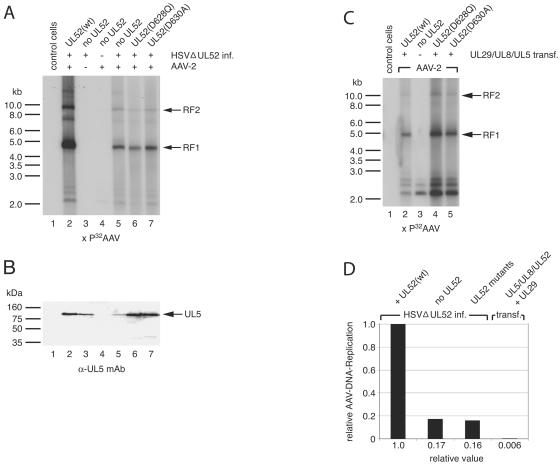
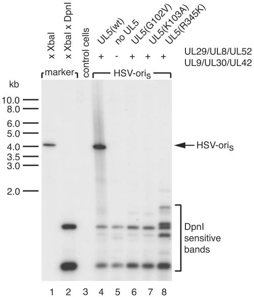
A subset of DNA replication proteins of herpes simplex virus (HSV) comprising the single-strand DNA-binding protein, ICP8 (UL29), and the helicase-primase complex (UL5, UL8, and UL52 proteins) has previously been shown to be sufficient for the replication of adeno-associated virus (AAV). We recently demonstrated complex formation between ICP8, AAV Rep78, and the single-stranded DNA AAV genome, both in vitro and in the nuclear HSV replication domains of coinfected cells. In this study the functional role(s) of HSV helicase and primase during AAV DNA replication were analyzed. To differentiate between their necessity as structural components of the HSV replication complex or as active enzymes, point mutations within the helicase and primase catalytic domains were analyzed. In two complementary approaches the remaining HSV helper functions were either provided by infection with HSV mutants or by plasmid transfection. We show here that upon cotransfection of the minimal four HSV proteins (i.e., the four proteins constituting the minimal requirements for basal AAV replication), UL52 primase catalytic activity was not required for AAV DNA replication. In contrast, UL5 helicase activity was necessary for fully efficient replication. Confocal microscopy confirmed that all mutants retained the ability to support formation of ICP8-positive nuclear replication foci, to which AAV Rep78 colocalized in a manner strictly dependent on the presence of AAV single-stranded DNA (ssDNA). The data indicate that recruitment of AAV Rep78 and ssDNA to nuclear replication sites by the four HSV helper proteins is maintained in the absence of catalytic primase or helicase activities and suggest an involvement of the HSV UL5 helicase activity during AAV DNA replication.


Figures

FIG. 1.
FIG. 1.
AAV ITR-dependent colocalization of Rep and ICP8 at nuclear HSV replication sites. HeLa cells were grown on glass coverslips and transfected with plasmids for the HSV helicase-primase complex (UL5, UL8, and UL52) and ICP8 (UL29). (A.I and A.II) Cells were cotransfected with an AAV wt plasmid. (B) Cells were transfected with a plasmid for an ITR-deleted AAV mutant lacking the terminal 145-bp ori sequences, required for the generation of AAV ssDNA. (C and D) Cells were cotransfected with plasmids for wt AAV (+ITR), ICP8, and the helicase-primase complex proteins omitting either UL5 (D) or UL52 (C) as indicated. Cells were fixed and stained with a polyclonal antibody reactive to Rep and a MAb reactive to ICP8 as outlined in Materials and Methods. Confocal microscopy for the detection of colocalization of ICP8 and Rep was conducted as outlined in results. The expression of Rep and ICP8 was detected by secondary antibodies conjugated to rhodamine (represented in red) or fluorescein (represented in green), respectively. Merged images of 0.6 μm cell slices reveal colocalization in yellow. Panel A.I shows images taken at a low cutoff value also applied in panels B to D. Images of the same cell taken at a higher cutoff value are presented in panel A.II to better reveal the complete colocalization of ICP8- and Rep-positive foci. Virtually no Rep fluorescence could be detected for the cells shown in panels B to D with the cutoff value chosen for panel A.II.
FIG. 2.
FIG. 2.
Role of the UL52 primase in AAV DNA replication. (A) HeLa cells were cotransfected with a plasmid for wt AAV-2 and expression constructs expressing either wt or mutated UL52 proteins as indicated. At 24 h posttransfection cells were infected with the UL52 null HSV mutant, hr114. Cells were harvested at 16 h postinfection, and low-molecular-weight DNA was extracted as outlined in Materials and Methods. Equal proportions of DNA samples were digested with DpnI to digest unreplicated input plasmids. DNA samples were fractionated on an agarose gel, blotted and hybridized to a 32P-labeled AAV probe. Typical AAV replicative intermediates appear at 4.7 kb as AAV monomer duplex form (RF1) and at 9.4 kb as AAV dimer duplex form (RF2), as indicated by the arrows on the right. (B) Western blot analysis of cell extracts of duplicate cell samples with mouse αUL5-MAb (MAb 376). The bands at 90 kDa represent UL5 helicase and are highlighted by an arrow. (C) HeLa cells were cotransfected with plasmids for wt AAV and expression constructs for ICP8 (UL29) and the helicase-primase complex, UL5, UL8, and UL52. In lanes 4 and 5, wt UL52 was replaced by the catalytic-site mutants, as indicated. Cells were harvested at 40 h posttransfection. AAV DNA replication was analyzed as outlined for panel A. (D) Quantification of the extent of AAV DNA replication initiated by HSV infection versus that upon transfection of the minimal set of HSV helper genes. AAV-transfected HeLa cells were infected with an HSV strain with a deletion of UL52 with [+UL52(wt) and UL52 mutants]) or without (no UL52) complementation by a UL52 expression plasmid. Cells were cotransfected with a plasmid for wt AAV and expression constructs for the four HSV helper proteins (UL5/UL8/UL52+ U29). Radioactive counts in the AAV replication intermediates RF1 and RF2 were quantified on the Southern blot by Phosphorimager analysis, as outlined in Materials and Methods. The counts for AAV replication in the presence of hr114 and the wt UL52 expression plasmid were set at 1.0, and the other values are displayed as fractions thereof.
FIG. 3.
FIG. 3.
HSV UL52 mutants are deficient in oriS-dependent HSV DNA replication. HeLa cells were cotransfected with a plasmid containing HSV-oriS (plasmid pH 10) and expression constructs for the seven HSV DNA replication proteins. In lanes 6 and 7 the wt UL52 plasmid was replaced by UL52 catalytic site mutants, as indicated. Total genomic DNA was extracted 40 h later and digested with XbaI and DpnI, as outlined in the legend of Fig. 2. A Southern blot was prepared and hybridized to a 32P-labeled HSV-oriS fragment. Replicated oriS is detected as a DpnI-resistant band of 4.3 kb, as indicated by the arrow. Plasmid pH 10 digested with either XbaI alone or with a combination of XbaI and DpnI served as markers run in parallel.
FIG. 4.
FIG. 4.
Colocalization of AAV Rep and HSV ICP8 is retained in the presence of HSV helicase or primase catalytic site mutants. The experiment was performed as outlined in the legend of Fig. 1. HeLa cells were grown on glass coverslips and transfected with plasmids encoding the HSV helicase-primase complex (UL5/8/52) and ICP8 (UL29). Panels A and G show wt HSV proteins, and in panels B to F and H to L, the wt HSV UL5 or UL52 proteins were replaced as indicated by catalytic site mutants. Plasmids specifying wt AAV or the ITR-deleted AAV mutant were also present in panels A to F and G to L, respectively. The anti-rabbit antibody reactive to Rep conjugated to rhodamine is shown in red and the anti-mouse antibody reactive to ICP8 conjugated to fluorescein is represented in green. Merged foci appear in yellow. A lower cutoff value was chosen for panels G to L compared to panels A to F to display the diffuse nuclear Rep staining pattern of low intensity in the absence of the AAV-ITRs.
FIG. 5.
FIG. 5.
The role of the UL5 helicase in AAV replication. (A) The experiment was performed as outlined in the legend of Fig. 2A, but the UL5 null mutant hr99 was used to initiate AAV DNA replication. Plasmids encoding wt UL5 or helicase motif I or IV mutants were used for complementation, as indicated. (B) A Western blot of protein samples prepared in parallel to the transfection experiment displayed in panel A was reacted with the mouse α-UL5 MAb 376 which was detected by enhanced chemiluminescence. The 90-kDa bands representing UL5 helicase are highlighted by an arrow. (C) The experiment was performed as described in the legend of Fig. 2C, but the wt UL5 plasmid was replaced by plasmids encoding the UL5 helicase motif mutants, as indicated. (D) Cell samples prepared in parallel to those in panel C were analyzed by Western blotting as described in the legend of panel B. (E) Quantification of AAV DNA replication upon hr99 infection in the presence of wt UL5, mutant UL5 or no UL5 plasmid compared to cotransfection of plasmids for wt AAV, ICP8, and the helicase-primase complex proteins (UL5/UL8/UL52+UL29). Phosphorimager analysis of AAV replication intermediates was performed as described in the legend of Fig. 2D.
FIG. 6.
FIG. 6.
HSV UL5 mutants are deficient in oriS-dependent HSV DNA replication. The experiment was performed essentially as described in the legend of Fig. 3 except that wt UL52 was used in all transfections and the wt UL5 plasmid was replaced by helicase motif mutants, as indicated.

Similar articles

Cited by

References

    1. Biswas, N., and S. K. Weller. 1999. A mutation in the C-terminal putative Zn2+ finger motif of UL52 severely affects the biochemical activities of the HSV-1 helicase-primase subcomplex. J. Biol. Chem. 274:8068-8076. - PubMed
    1. Biswas, N., and S. K. Weller. 2001. The UL5 and UL52 subunits of the herpes simplex virus type 1 helicase-primase subcomplex exhibit a complex interdependence for DNA binding. J. Biol. Chem. 276:17610-17619. - PubMed
    1. Carrington-Lawrence, S. D., and S. K. Weller. 2003. Recruitment of polymerase to herpes simplex virus type 1 replication foci in cells expressing mutant primase (UL52) proteins. J. Virol. 77:4237-4247. - PMC - PubMed
    1. Chen, Y., S. D. Carrington-Lawrence, P. Bai, and S. K. Weller. 2005. Mutations in the putative zinc-binding motif of UL52 demonstrate a complex interdependence between the UL5 and UL52 subunits of the human herpes simplex virus type 1 helicase/primase complex. J. Virol. 79:9088-9096. - PMC - PubMed
    1. Chiorini, J. A., S. M. Wiener, R. A. Owens, S. R. M. Kyöstiö, R. M. Kotin, and B. Safer. 1994. Sequence requirements for stable binding and function of Rep68 on the adeno-associated virus type 2 inverted terminal repeats. J. Virol. 68:7448-7457. - PMC - PubMed

Publication types

LinkOut - more resources