. 2006 Jun;80(11):5308-20.
doi: 10.1128/JVI.02460-05.

Characterization of the early steps of hepatitis C virus infection by using luciferase reporter viruses

Affiliations

Characterization of the early steps of hepatitis C virus infection by using luciferase reporter viruses

George Koutsoudakis et al. J Virol. 2006 Jun.

Abstract

The lack of an efficient system to produce hepatitis C virus (HCV) particles has impeded the analysis of the HCV life cycle. Recently, we along with others demonstrated that transfection of Huh7 hepatoma cells with a novel HCV isolate (JFH1) yields infectious viruses. To facilitate studies of HCV replication, we generated JFH1-based bicistronic luciferase reporter virus genomes. We found that RNA replication of the reporter construct was only slightly attenuated and that virus titers produced were only three- to fivefold lower compared to the parental virus, making these reporter viruses an ideal tool for quantitative analyses of HCV infections. To expand the scope of the system, we created two chimeric JFH1 luciferase reporter viruses with structural proteins from the Con1 (genotype 1b) and J6CF (genotype 2a) strains. Using these and the authentic JFH1 reporter viruses, we analyzed the early steps of the HCV life cycle. Our data show that the mode of virus entry is conserved between these isolates and involves CD81 as a key receptor for pH-dependent virus entry. Competition studies and time course experiments suggest that interactions of HCV with cell surface-resident glycosaminoglycans aid in efficient infection of Huh7 cells and that CD81 acts during a postattachment step. The reporter viruses described here should be instrumental for investigating the viral life cycle and for the development of HCV inhibitors.


Figures
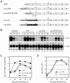
FIG. 1.
FIG. 1.
Genetic organization and replication of JFH1 and JFH1-derived luciferase reporter constructs. (A) A schematic representation of parental JFH1 genome is given at the top, and derivates with the reporter gene used in this study are shown below. The 5′ and 3′ NTR are drawn as thick black lines, and the JFH1 polyprotein is depicted as an open box; individual proteins are separated by vertical lines. In the case of reporter constructs, viral proteins are expressed via an internal EMCV IRES (gray bar). Note that Luc-JFH1/ΔE1-E2 carries a large in frame deletion in the coding region for E1-E2, which is known to inactivate virus particle formation (51). The luciferase gene (hatched) is fused in frame to the 16 N-terminal residues of the JFH1 core coding region (white). Chimeric genomes are depicted with J6CF (GT 2a) and Con1 (GT 1b) proteins in gray and black boxes, respectively. The junction between the different isolates is located within NS2. (B) RNA replication of JFH1, Luc-JFH1, and Luc-JFH1/ΔE1-E2 as determined by Northern hybridization. In vitro transcribed RNAs of given constructs were transfected into Huh7-Lunet cells that were harvested at the time points specified above the panel. Total RNA was prepared and analyzed with an HCV-specific probe. Equal sample loading was monitored by detection of β-actin RNA (bottom). As controls, total RNA of naive Huh7-Lunet cells (N) and serial dilutions of in vitro transcribed RNAs spiked into total RNA of naïve cells were loaded in parallel (lanes 1 to 4). (C) Quantification of the Northern blot shown in panel B by phosphor imaging. The amount of HCV RNA/μg of total RNA was calculated by comparison with the controls loaded in parallel. Values were corrected for differences in loading by using β-actin signals. (D) Luciferase activity in transiently transfected Huh7-Lunet cells. Transfected cells were lysed at given time points, and the amount of luciferase activity was determined and is given in relative light units (RLU) per well (each point is the average value of duplicate wells; the error bars show standard deviations of the means).
FIG. 2.
FIG. 2.
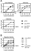
Release of core protein and infectivity upon transfection of JFH1 constructs into Huh7-Lunet cells. (A) In vitro transcribed RNAs of JFH1 (black circles), Luc-JFH1 (black diamonds), and Luc-JFH1ΔE1-E2 (open squares) were transfected into Huh7-Lunet cells, and cell-free culture fluids were collected at 4 h to 96 h posttransfection. The amount of core protein present in the respective culture medium was determined employing a core-specific immunoassay (upper left graph). In parallel, supernatants were used to inoculate naïve Huh7-Lunet cells, and infectivity associated with the respective samples was quantified by using an immunofluorescence-based limiting dilution assay (upper right graph) and (where applicable) by assessing the amount of reporter activity present in target cells 72 h postinoculation (lower left graph). The gray lines indicate the background of the reporter assay from naïve Huh7-Lunet cells and the detection limit of the core ELISA, respectively. Mean values of two experiments are given; error bars represent standard errors of the means. (B) Huh7-Lunet cells were inoculated with serial dilutions of Luc-JFH1-containing medium or with undiluted Luc-JFH1/ΔE1-E2 culture fluid and lysed at the indicated time points postinoculation to quantify luciferase activity. Mean values of two independent wells are given (error bars show standard deviations). The gray line indicates the background of the assay determined by measuring mock-infected cells.
FIG. 3.
FIG. 3.
Influence of cell growth on replication and release of infectious Luc-JFH1 virus particles. Huh7-Lunet cells were transfected with Luc-JFH1 RNA and seeded into multiple culture dishes with the given cell density (8.32 × 10⁴ to 0.52 × 10⁴ cells/cm2). Luciferase activity in transfected cells was determined at the indicated time points posttransfection (left). Cell-free supernatants from cells seeded with the given densities were collected 24 to 144 h after transfection and used to inoculate naïve Huh7-Lunet cells. Luciferase activity in the inoculated cells was determined 72 h later (right). Mean values of two independent wells measured are given (error bars represent the standard errors of the means).
FIG. 4.
FIG. 4.
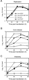
Replication and production of infectious virus particles upon transfection of chimeric reporter viruses. (A) Replication kinetics of given HCV genomes and the deletion mutant as determined by luciferase assay measured at various time points posttransfection. Release of core protein (B) and infectivity into the supernatant (C) were quantified by using a core-specific immunoassay and by inoculating naïve Huh7-Lunet cells and subsequent luciferase assays, respectively. ELISAs were performed in singleton; for reporter assays, two independent wells were analyzed, and mean values are given (error bars represent the standard errors of the means).
FIG. 5.
FIG. 5.
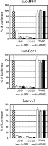
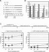
Neutralization of genotype 1b and 2a HCV infection by different CD81-specific antibodies. Huh7-Lunet cells were infected with reporter viruses in the presence of increasing amounts of CD81-specific (JS-81 and 1.3.3.22) antibodies or isotype-matched control antibodies (WM15) directed against CD13, an irrelevant surface molecule on Huh7-Lunet cells. Inoculation was terminated after 4 h by washing cells with PBS and supplementing them with fresh culture medium without antibodies. Luciferase activity was determined 72 h postinfection and is expressed relative to the amount observed in the absence of inhibitory antibodies (hatched bar). White, gray, and black bars represent infections in the presence of antibodies at a concentration of 0.08 μg/ml, 0.4 μg/ml, and 2 μg/ml, respectively. The graphs (from top to bottom) depict results obtained with Luc-JFH1, Luc-Con1, and Luc-Jc1 particles, respectively. Mean values of duplicate wells are given (error bars represent standard errors of the means).
FIG. 6.
FIG. 6.
Pretreatment of target cells with entry inhibitors. Huh7-Lunet cells were incubated with regular medium or medium supplemented with heparin (50 μg/ml) or antibodies against CD81 (JS-81; 2 μg/ml) for 1 h at 4°C. Subsequently, cells were washed with PBS and inoculated with reporter viruses. Infection was quantified as described in the legend of Fig. 5.
FIG. 7.
FIG. 7.
Infection of Huh7-Lunet cells after pretreatment with GAG-lyases. Huh7-Lunet cells were treated with buffer alone (mock) or with buffer containing 0.5 U/ml heparinase I, heparinase III, or chondroitinase ABC for 1 h at 37°C. Subsequently, cells were washed three times with PBS and then inoculated with Luc-Jc1 for 1 h at 37°C. The efficiency of infection was determined 48 h postinoculation as described in the legend of Fig. 5.
FIG. 8.
FIG. 8.
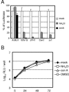
Susceptibility of HCV entry to drugs inhibiting endosomal acidification. (A) Huh7-Lunet cells were mock-treated (open bars) or incubated for 1 h with medium containing 10 mM NH4Cl or 5 nM ConA (gray or black bars, respectively), washed with medium, and infected with different viruses for 4 h in the presence or absence of the drugs. As control for pH-dependent or -independent virus entry, infections with retroviral pseudo-particles bearing VSV-G or A-MLV envelope proteins, respectively, were performed in the same way. Luciferase activity was determined 72 h postinfection and is expressed relative to the values obtained in the absence of drugs. Mean values of duplicates and the standard errors of the means are given. (B) ConA and NH4Cl do not interfere with HCV RNA replication. Huh7-Lunet cells carrying selectable JFH1-based luciferase reporter replicons were seeded into replicate wells. At 24 h after seeding, cells were treated with drugs for 5 h. RNA replication was monitored by measuring reporter activity at the given time points posttreatment. Mean values of duplicates and the standard errors of the means are given.
FIG. 9.
FIG. 9.
Kinetics of inhibitory activity exerted by various compounds. (A) Schematic drawing of the experimental setup of the experiment depicted in panel B. Inhibition of Luc-Jc1 entry into Huh7-Lunet cells by heparin (50 μg/ml), CD81-specific antibodies (JS-81; 2 μg/ml) and ConA (5 nM) was compared using five different experimental protocols (indicated by roman numerals I through V). Virus binding to target cells was performed for 1 h at 4°C in the absence (II to IV) or in the presence (I) of compounds. Subsequently, cells were washed with PBS and shifted to 37°C to allow entry to proceed. Depending on the protocol, inhibitors were added directly or 1, 2 or 3 h thereafter (II, III, IV, and V, respectively). Dotted lines indicate the time interval during which an inhibitor was present; black arrows indicate the addition and removal of virus inoculum (B). Efficiency of infection using the compounds given below the bars and the protocols described for panel A was determined by luciferase assays 72 h postinfection and is expressed relative to infection in the absence of inhibitors (black bar). Mean values of two wells and the standard errors of the means are given. (C) A schematic drawing of the experimental setup is depicted at the top of the panel. Huh7-Lunet cells were incubated with Luc-Jc1 virus for various times at 37°C in the presence of heparin (50 μg/ml; left) or anti-CD81 antibodies (2 μg/ml; right) or in the absence of either compound (black diamonds and open squares, respectively). Subsequently, virus inocula were removed; cells were washed with PBS and supplemented with fresh culture medium containing either heparin (50 μg/ml) or anti-CD81 antibodies (2 μg/ml). Four hours later, culture medium was replaced by medium without inhibitors. The efficiency of infection was measured by luciferase assays 48 h later and is given for each time point relative to control infections performed in the same way but always without inhibitor. Mean values of two wells and the standard errors of the means are given.

References

    1. Agnello, V., G. Abel, M. Elfahal, G. B. Knight, and Q. X. Zhang. 1999. Hepatitis C virus and other Flaviviridae viruses enter cells via low density lipoprotein receptor. Proc. Natl. Acad. Sci. USA 96:12766-12771. - PMC - PubMed
    1. Bartenschlager, R., M. Frese, and T. Pietschmann. 2004. Novel insights into hepatitis C virus replication and persistence. Adv. Virus Res. 63:71-180. - PubMed
    1. Barth, H., C. Schafer, M. I. Adah, F. Zhang, R. J. Linhardt, H. Toyoda, A. Kinoshita-Toyoda, T. Toida, T. H. Van Kuppevelt, E. Depla, F. von Weizsacker, H. E. Blum, and T. F. Baumert. 2003. Cellular binding of hepatitis C virus envelope glycoprotein E2 requires cell surface heparan sulfate. J. Biol. Chem. 278:41003-41012. - PubMed
    1. Bartosch, B., J. Dubuisson, and F. L. Cosset. 2003. Infectious hepatitis C virus pseudo-particles containing functional E1-E2 envelope protein complexes. J. Exp. Med. 197:633-642. - PMC - PubMed
    1. Bartosch, B., A. Vitelli, C. Granier, C. Goujon, J. Dubuisson, S. Pascale, E. Scarselli, R. Cortese, A. Nicosia, and F. L. Cosset. 2003. Cell entry of hepatitis C virus requires a set of co-receptors that include the CD81 tetraspanin and the SR-B1 scavenger receptor. J. Biol. Chem. 278:41624-41630. - PubMed

Publication types