. 2006 Jun;80(11):5499-508.
doi: 10.1128/JVI.00200-06.

Prediction and identification of herpes simplex virus 1-encoded microRNAs

Affiliations

Prediction and identification of herpes simplex virus 1-encoded microRNAs

Can Cui et al. J Virol. 2006 Jun.

Abstract

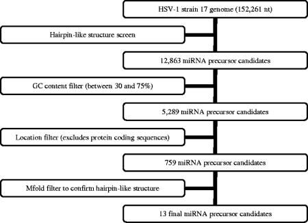
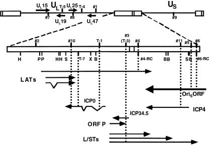
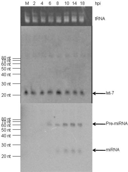
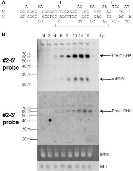
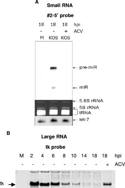
MicroRNAs (miRNAs) are key regulators of gene expression in higher eukaryotes. Recently, miRNAs have been identified from viruses with double-stranded DNA genomes. To attempt to identify miRNAs encoded by herpes simplex virus 1 (HSV-1), we applied a computational method to screen the complete genome of HSV-1 for sequences that adopt an extended stem-loop structure and display a pattern of nucleotide divergence characteristic of known miRNAs. Using this method, we identified 11 HSV-1 genomic loci predicted to encode 13 miRNA precursors and 24 miRNA candidates. Eight of the HSV-1 miRNA candidates were predicted to be conserved in HSV-2. The precursor and the mature form of one HSV-1 miRNA candidate, which is encoded approximately 450 bp upstream of the transcription start site of the latency-associated transcript (LAT), were detected during infection of Vero cells by Northern blot hybridization. These RNAs, which behave as late gene products, are not predicted to be conserved in HSV-2. Additionally, small RNAs, including some that are roughly the expected size of precursor miRNAs, were detected using probes for miRNA candidates derived from sequences encoding the 8.3-kilobase LAT, from sequences complementary to U(L)15 mRNA, and from the region between ICP4 and U(S)1. However, no species the size of typical mature miRNAs were detected using these probes. Three of these latter miRNA candidates were predicted to be conserved in HSV-2. Thus, HSV-1 encodes at least one miRNA. We hypothesize that HSV-1 miRNAs regulate viral and host gene expression.


Figures

FIG. 1.
FIG. 1.
Flowchart of the HSV-1 miRNA prediction procedure. See the text for details.
FIG. 2.
FIG. 2.
Genomic positions of predicted HSV-1 miRNA precursors. The HSV-1 genome in the prototype orientation is shown at the top. UL and US denote the unique sequences of the long (L) and short (S) components of the genome, respectively, which are labeled with solid lines, and the open boxes denote repeat sequences. The L-S junction region is expanded below, with restriction endonuclease cleavage sites abbreviated as follows: B, BamHI; H, HpaI; P, PstI; S, SalI; and X, XhoI. Locations and orientations of transcripts in this region are denoted by solid arrows, with angled lines denoting portions removed by splicing. Numbers indicate locations of predicted miRNA precursors in the viral genome, with those in UL and US shown on the top and those in the repeat sequences below. Those above the line or box are predicted to be transcribed from left to right, while those below indicate the opposite direction of transcription. Dotted lines indicate positions of the predicted miRNA precursors relative to different transcripts. In the unique sequences, only transcripts antisense to predicted miRNA precursors are shown. See Table 1 for exact genomic coordinates of the predicted HSV-1 miRNA precursors.
FIG. 3.
FIG. 3.
Analysis of HSV-1 miRNAs. Small RNAs harvested from mock-infected (M) or from HSV-1-infected cells at the times indicated at the top of the figure were separated by PAGE, stained for tRNA with ethidium bromide (top panel), and blotted to a membrane for hybridization with a probe for let-7 miRNA (middle panel) or a pool of probes for predicted HSV-1 miRNAs (bottom panel). This pool contained probes 1-5′, 2-5′, 3-5′, 4-5′, 5-5′, 6-5′, 7-5′, 8, 9-5′, and 11-5′. The position of tRNA is shown to the right of the top panel. The sizes of RNA markers are indicated to the left of the middle and the bottom panels. The positions of let-7 miRNA, HSV-1 miRNAs, and their precursors are indicated to the right of the middle and the bottom panels, respectively.
FIG. 4.
FIG. 4.
Identification of an HSV-1 miRNA. (A) The sequence of predicted HSV-1 miRNA precursor 2 is shown. The upper strand is the 5′ strand, and the lower strand is the 3′ strand. The predicted mature miRNAs, which were used to design probes, are indicated with brackets. (B) Northern blot analysis of predicted HSV-1 miRNA precursor 2 using small RNAs harvested from mock-infected (M) or HSV-1-infected cells at the times indicated at the top of the figure. The top panel shows a phosphorimage of the pattern of hybridization using the probe to the 5′ strand of predicted precursor 2, and the next panel shows the pattern using the probe to the 3′ strand. The third panel shows a photograph under UV light of a portion of the ethidium bromide-stained gel prior to membrane transfer, and the bottom panel shows a phosphorimage of the pattern of hybridization using a let-7 probe. The sizes of RNA markers are indicated to the left of the top and the second panels. The positions of HSV-1 pre-miRNA, miRNA, tRNA, and let-7 are indicated to the right of the figure.
FIG. 5.
FIG. 5.
Effect of acyclovir (ACV) on HSV-1 miR-H1 expression. (A) Small RNAs were harvested from mock-infected (M) or from HSV-1-infected cells in the absence (−) or presence (+) of ACV at 18 h. The top panel shows a phosphorimage of the pattern of hybridization with probe 2-5′. The middle panel shows a photograph under UV of a portion of ethidium bromide-stained gel prior to membrane transfer. The bottom panel shows a phosphorimage of the pattern of hybridization using a let-7 probe. The positions of pre-miR-H1 (pre-miR), miR-H1 (miR), 5.8S rRNA, 5S rRNA, tRNA, and let-7 are indicated to the right of the figure. (B) Large RNAs were harvested from mock-infected (M) or from HSV-1-infected cells in the absence (−) or presence (+) of ACV at the times indicated at the top of the figure, separated by agarose gel electrophoresis, and blotted to membrane for hybridization with a tk probe. The position of tk mRNA is indicated to the left of the phosphorimage.
FIG. 6.
FIG. 6.
Identification of small RNA species specific to HSV-1 infection. Small RNAs harvested from mock-infected (M) or from HSV-1-infected cells at the times indicated at the top of the figure were separated by PAGE, stained with ethidium bromide (UV visualization of a portion of the gel is shown in the third gel in panel A and in the second gels in panels B, C, and D), and blotted to membrane for hybridization with 4-5′ probe (A, top gel), 4-3′ probe (A, second gel), 6-RC probe (B, top gel), 7-3′ probe (C, top gel), and T-1 probe (D, top gel). The sizes of RNA markers are indicated to the left of the phosphorimages. The position of tRNA and the putative pre-miRNAs are indicated to the right of the figure. (A to C) The membrane was stripped and hybridized with a probe to let-7, the position of which is also shown to the right of the bottom gels.

Similar articles

Cited by

References

    1. Altschul, S. F., T. L. Madden, A. A. Schaffer, J. Zhang, Z. Zhang, W. Miller, and D. J. Lipman. 1997. Gapped BLAST and PSI-BLAST: a new generation of protein database search programs. Nucleic Acids Res. 25:3389-3402. - PMC - PubMed
    1. Ambros, V. 2004. The functions of animal microRNAs. Nature 431:350-355. - PubMed
    1. Ambros, V., B. Bartel, D. P. Bartel, C. B. Burge, J. C. Carrington, X. Chen, G. Dreyfuss, S. R. Eddy, S. Griffiths-Jones, M. Marshall, M. Matzke, G. Ruvkun, and T. Tuschl. 2003. A uniform system for microRNA annotation. RNA 9:277-279. - PMC - PubMed
    1. Baines, J. D., C. Cunningham, D. Nalwanga, and A. Davison. 1997. The UL15 gene of herpes simplex virus type 1 contains within its second exon a novel open reading frame that is translated in frame with the UL15 gene product. J. Virol. 71:2666-2673. - PMC - PubMed
    1. Bartel, D. P. 2004. MicroRNAs: genomics, biogenesis, mechanism, and function. Cell 116:281-297. - PubMed

Publication types