. 2006 Jun;80(11):5531-9.
doi: 10.1128/JVI.02725-05.

A herpes simplex virus recombinant that exhibits a single-chain antibody to HER2/neu enters cells through the mammary tumor receptor, independently of the gD receptors

Affiliations

A herpes simplex virus recombinant that exhibits a single-chain antibody to HER2/neu enters cells through the mammary tumor receptor, independently of the gD receptors

Laura Menotti et al. J Virol. 2006 Jun.

Abstract

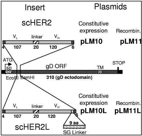
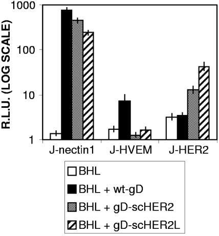
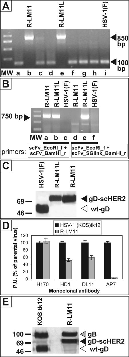
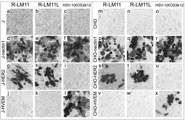
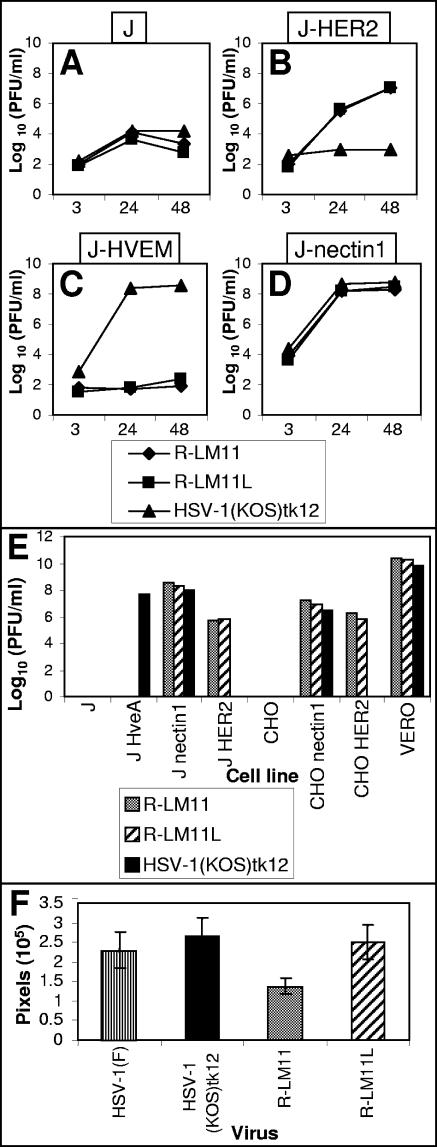
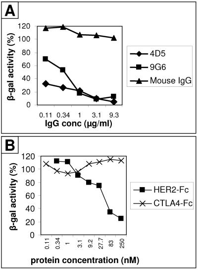
The human epidermal growth factor receptor 2/neuregulin (HER2/neu) receptor is overexpressed in highly malignant mammary and ovarian tumors and correlates with a poor prognosis. It is a target for therapy; humanized monoclonal antibodies to HER2 have led to increased survival of patients with HER2/neu-positive breast cancer. As a first step in the design of an oncolytic herpes simplex virus able to selectively infect HER2/neu-positive cells, we constructed two recombinants, R-LM11 and R-LM11L, that carry a single-chain antibody (scFv) against HER2 inserted at residue 24 of gD. The inserts were 247 or 256 amino acids long, and the size of the gD ectodomain was almost doubled by the insertion. We report the following. R-LM11 and R-LM11L infected derivatives of receptor-negative J or CHO cells that expressed HER2/neu as the sole receptor. Entry was dependent on HER2/neu, since it was inhibited in a dose-dependent manner by monoclonal antibodies to HER2/neu and by a soluble form of the receptor. The scFv insertion in gD disrupted the ability of the virus to enter cells through HVEM but maintained the ability to enter through nectin1. This report provides proof of principle that gD can tolerate fusion to a heterologous protein almost as large as the gD ectodomain itself without loss of profusion activity. Because the number of scFv's to a variety of receptors is continually increasing, this report makes possible the specific targeting of herpes simplex virus to a large collection of cell surface molecules for both oncolytic activity and visualization of tumor cells.


Figures

FIG. 1.
FIG. 1.
Schematic representation of the gD gene carrying the sequence encoding scHER2. The cDNA encoding scHER2 was inserted into the gD gene, previously modified to carry EcoRI and BamHI restriction sites. The insert was flanked by sequences encoding 4 and 6 aa residues at the 5′ and 3′ ends, respectively, to restore the complete gD sequence. pLM10L and pLM11L carried a 9-aa Ser-Gly linker (L) downstream of the insert. In pLM10 and pLM10L, the chimeric gD gene was cloned into pcDNA3.1 for constitutive expression. pLM11 and pLM11L carry the chimeric gD gene bracketed by upstream and downstream gD sequences and were employed to generate recombinant (Recombin.) viruses. Numbers indicate the length in amino acid residues of each fragment. The insertion of the EcoRI site in gD caused the D21G and L22I substitutions in mature gD. VH and VL, heavy- and light-chain variable domains of the anti-HER2/neu antibody 4D5. sp, signal peptide. Bars are drawn to scale.
FIG. 2.
FIG. 2.
Expression of HER2/neu in the stable clonal cell lines J-HER2 (A) and CHO-HER2 (B). Cells were fixed with paraformaldehyde and reacted with MAb 9G6 to the HER2 ectodomain, followed by an FITC-conjugated secondary antibody. Fluorescence localizes at the plasma membrane.
FIG. 3.
FIG. 3.
Cell-cell fusion mediated by chimeric scHER2-gD. The effector COS cells, cotransfected with plasmids encoding gB, gH, gL (BHL), and pCAGT7pol plus chimeric or wt gD, were cocultivated with J-nectin1, J-HVEM, or J-HER2 target cells transfected with pEMCVLuc. Luciferase activity was expressed as relative light units (RLU) on a log scale. Each experiment was performed at least three times, and samples were run in triplicate; mean values are shown. Vertical bars, standard deviations.
FIG. 4.
FIG. 4.
Recombinant viruses harbor the scFv to HER2 in gD and express a chimeric gD. (A and B) Amplification of the sequences encoding scHER2 from lysates of R-LM11- and R-LM11L-infected cells. (A) PCR was performed with primers annealing to the gD sequences that flank the site for scHER2 insertion. The presence of the insert causes an increase in the size of the amplification product from 100 bp (nonrecombinant plaques [lanes a, c, d, f, g, and h]) to 850 bp (lanes b and e). Lane i, HSV-1(F), used as a control. MW, 1-kb DNA ladder. (B) PCR was performed with primers annealing to the scHER2 insert. Lanes a to c, PCR with primers used for cloning the scHER2 insert. Lanes d to f, PCR with the same forward primer as in lanes a to c and a reverse primer annealing to the serine-glycine linker, thus amplifying R-LM11L rather than R-LM11. The amplification products from the recombinants exhibit the expected length (∼750 bp). HSV-1(F) was used as a negative control and did not give rise to any amplification product (lanes c and f). (C) Electrophoretic mobility of chimeric scHER2-gD. Lysates of cells infected with R-LM11, R-LM11L, or HSV-1(F) were subjected to SDS-PAGE, transferred to nitrocellulose membranes, and visualized by Western blotting with MAb H170 against gD, followed by peroxidase-conjugated anti-mouse IgG and ECL. In the recombinants, the presence of scHER2 results in a slower-migrating band (black arrowhead) than that with wt gD (white arrowhead). Numbers to the left represent migration positions of molecular mass markers (in kilodaltons). (D) Reactivities of wt gD and the gD-HER2 chimera to a panel of monoclonal antibodies, measured by cell ELISA (21). Vero cells were infected with the indicated viruses. At 16 h after infection, they were reacted with the indicated antibodies, followed by a peroxidase-conjugated anti-mouse antibody and o-phenylenediamine. Binding of antibodies was quantified as peroxidase units (P.U.), and expressed as a percentage relative to the cells infected with the parental HSV-1(KOS)tk12 virus. Each assay was performed in quadruplicate. Bars represent means. Error bars, standard deviations. (E) Quantification of gD and gB present in virions. Virions were pelleted from the extracellular medium of infected Vero cells after growth for 24 h. Equal amounts of virions, measured as PFU, were loaded for SDS-PAGE separation. Amounts of gB and gD were detected by Western blotting with MAbs H1817 and H170, respectively. Arrowheads indicate migration positions.
FIG. 5.
FIG. 5.
The recombinant viruses R-LM11 and R-LM11L infect cells via the HER2/neu receptor. Micrographs show J (a to c), J-nectin1 (d to f), J-HER2 (g to i), J-HVEM (j to l), CHO (m to o), CHO-nectin1 (p to r), CHO-HER2 (s to u), and CHO-HVEM (v to x) cells exposed to the recombinant virus R-LM11 (a, d, g, j, m, p, s, and v) or R-LM11L (b, e, h, k, n, q, t, and w) or to the parental virus HSV-1(KOS)tk12 (c, f, i, l, o, r, u, and x) at 10 PFU/cell. Infection was monitored as β-galactosidase activity by X-Gal staining 16 h following infection.
FIG. 6.
FIG. 6.
Growth and plaque formation of R-LM11 and R-LM11L recombinants. (A to D) Growth curves of R-LM11 and R-LM11L. Replicate cultures of J (A), J-HER2 (B), J-HVEM (C), or J-nectin1 (D) cells were infected with R-LM11 (♦), R-LM11L (▪), or HSV-1(KOS)tk12 (▴) at 10 PFU/cell. Progeny virus was harvested at 3, 24, or 48 h after infection and titrated on Vero cells. (E) Plaque formation of R-LM11 and R-LM11L. R-LM11 (gray bars), R-LM11L (hatched bars), and HSV-1(KOS)tk12 (black bars) were plated in the indicated cell lines. Monolayers were fixed at 24 or 48 h after infection, and plaques were visualized by X-Gal or Giemsa staining. (F) Plaques formed as shown in panel E were photographed, and the plaque areas were measured by means of the Histogram program and expressed as pixels. For each virus, the areas of at least 20 plaques were measured. Histograms represent averages; error bars, standard deviations.
FIG. 7.
FIG. 7.
Infection of R-LM11 and R-LM11L recombinants in HER2/neu-expressing cells is blocked by antibodies to HER2/neu and by a soluble form of HER2, HER2-Fc. (A) J-HER2 cells were preincubated with the indicated concentrations of purified IgG of monoclonal antibody 4D5 (♦) or 9G6 (▪) against HER2/neu or of irrelevant mouse IgGs (▴) for 1 h at 37°C. Virus was added to the antibody-containing medium and allowed to infect the cells for 90 min at 37°C. Infection was monitored 16 h later as β-galactosidase activity. (B) Replicate aliquots of R-LM11L were preincubated with the indicated concentrations of purified soluble recombinant HER2-Fc (▪) or CTLA4-Fc (×) for 1 h at 37°C and allowed to absorb to CHO-HER2 cells for 90 min at 37°C. Infection was quantified 16 h later as β-galactosidase activity. Each point represents the average of triplicate assays. The standard error ranged from 0.6 to 1.9% of mean values. One hundred percent indicates the optical density measured in untreated, virus-infected cultures.

References

    1. Advani, S. J., R. R. Weichselbaum, R. J. Whitley, and B. Roizman. 2002. Friendly fire: redirecting herpes simplex virus-1 for therapeutic applications. Clin. Microbiol. Infect. 8:551-563. - PubMed
    1. Andreansky, S., L. Soroceanu, E. R. Flotte, J. Chou, J. M. Markert, G. Y. Gillespie, B. Roizman, and R. J. Whitley. 1997. Evaluation of genetically engineered herpes simplex viruses as oncolytic agents for human malignant brain tumors. Cancer Res. 57:1502-1509. - PubMed
    1. Avitabile, E., G. Lombardi, and G. Campadelli-Fiume. 2003. Herpes simplex virus glycoprotein K, but not its syncytial allele, inhibits cell-cell fusion mediated by the four fusogenic glycoproteins, gD, gB, gH, and gL. J. Virol. 77:6836-6844. - PMC - PubMed
    1. Bergman, I., P. Whitaker-Dowling, Y. Gao, and J. A. Griffin. 2004. Preferential targeting of vesicular stomatitis virus to breast cancer cells. Virology 330:24-33. - PubMed
    1. Brandimarti, R., T. Huang, B. Roizman, and G. Campadelli Fiume. 1994. Mapping of herpes simplex virus 1 genes with mutations which overcome host restrictions to infection. Proc. Natl. Acad. Sci. USA 91:5406-5410. - PMC - PubMed

Publication types