. 2006 Jun;80(11):5588-98.
doi: 10.1128/JVI.00060-06.

Human cytomegalovirus attenuates interleukin-1beta and tumor necrosis factor alpha proinflammatory signaling by inhibition of NF-kappaB activation

Affiliations

Human cytomegalovirus attenuates interleukin-1beta and tumor necrosis factor alpha proinflammatory signaling by inhibition of NF-kappaB activation

Michael A Jarvis et al. J Virol. 2006 Jun.

Abstract

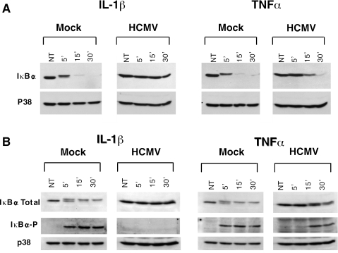
Viral infection is associated with a vigorous inflammatory response characterized by cellular infiltration and release of the proinflammatory cytokines interleukin-1 (IL-1) and tumor necrosis factor alpha (TNF-alpha). In the present study, we identified a novel function of human cytomegalovirus (HCMV) that results in inhibition of IL-1 and TNF-alpha signaling pathways. The effect on these pathways was limited to cells infected with the virus, occurred at late times of infection, and was independent of cell type or virus strain. IL-1 and TNF-alpha signaling pathways converge at a point upstream of NF-kappaB activation and involve phosphorylation and degradation of the NF-kappaB inhibitory molecule IkappaBalpha. The HCMV inhibition of IL-1 and TNF-alpha pathways corresponded to a suppression of NF-kappaB activation. Analysis of IkappaBalpha phosphorylation and degradation suggested that HCMV induced two independent blocks in NF-kappaB activation, which occurred upstream from the point of convergence of the IL-1 and TNF-alpha pathways. We believe that the ability of HCMV to inhibit these two major proinflammatory pathways reveals a critical aspect of HCMV biology, with possible importance for immune evasion, as well as establishment of infection in cell types persistently infected by this virus.


Figures

FIG. 1.
FIG. 1.
qRT-PCR showing that HCMV inhibits IL-1β- and TNF-α-mediated induction of multiple inflammatory genes. HFF cells were either infected with HCMV TR at an MOI of 3 (black bars) or mock infected (white bars). At day 3 p.i., cells were treated with IL-1β (1 ng/ml) or TNF-α (10 ng/ml). At 1 and 3 h posttreatment, cells were harvested for RNA extraction and preparation of cDNA. Levels of chemokine expression (IL-8, IL-6, MCP-1, and GRO-α) were measured by qRT-PCR. Chemokine induction was measured by absolute quantification and then normalized to GAPDH levels to determine induction (n-fold). The results shown are representative of two independent experiments.
FIG. 2.
FIG. 2.
ELISA showing HCMV inhibition of IL-1 and TNF-α proinflammatory pathways corresponds to reduced release of IL-8. HFF cells and endothelial cells (HUVECs) were infected with HCMV TR at an MOI of 3 (black bars) or mock infected (white bars), followed by treatment with IL-1β or TNF-α at day 3 p.i. After 6 h, supernatants were harvested, clarified by centrifugation, and stored at −80°C prior to analysis. The concentration of IL-8 released from cells was determined by ELISA. Levels of released IL-8 were determined by comparison to a standard curve and are presented as percentages of IL-8 levels released from noninduced cells.
FIG. 3.
FIG. 3.
HCMV inhibition of IL-1 and TNF-α proinflammatory pathways occurs at later times of infection and is independent of virus strain. (A) Endothelial cells (HUVECs) were infected with HCMV TR, or (B) HFF cells were infected with either HCMV AD169 or HCMV Towne at an MOI of 0.5. At day 3 p.i., cells were treated with IL-1β. After 6 h, cells were fixed and incubated with antibodies against the HCMV IE protein IE2 (Cy5; blue), the E/L protein gB (fluorescein isothiocyanate; green), and cellular IL-8 (Texas Red; red). Immunoreactivity was detected with the appropriate fluorophore-conjugated secondary Ab and visualized by confocal microscopy.
FIG. 4.
FIG. 4.
HCMV inhibition of IL-1 pathway corresponds to an inhibition of NF-κB activation. Endothelial cells (tAEC-5A) were infected with HCMV TR at an MOI of 0.5. At day 3 p.i., cells were treated with IL-1β or untreated. After 30 min, cells were fixed and incubated with Abs against the HCMV IE protein IE2 (Texas Red; red), the E/L protein gB (fluorescein isothiocyanate; green), and cellular NF-κB (Cy5; blue). Immunoreactivity was detected with the appropriate fluorophore-conjugated secondary Ab and visualized by confocal microscopy.
FIG. 5.
FIG. 5.
HCMV inhibition of NF-κB activation is distinct between IL-1 and TNF-α pathways. HFF cells were infected with HCMV TR at an MOI of 0.5. At day 3 p.i., cells were treated with IL-1β or TNF-α. After 30 min, cells were fixed and incubated with antibodies against the HCMV E/L protein gB (fluorescein isothiocyanate; green) and cellular NF-κB (Cy5; blue). Immunoreactivity was detected with the appropriate fluorophore-conjugated secondary Ab and visualized by confocal microscopy.
FIG. 6.
FIG. 6.
Analysis of IκBα degradation shows that HCMV induces a differential inhibition of IL-1 signaling pathways compared to that of TNF-α signaling pathways. HFF cells were infected with HCMV TR at an MOI of 3 or mock infected. At day 3 p.i., cells were treated with IL-1β, TNF-α, or dimethyl sulfoxide (diluent control). (A) At increasing times postinduction, cells were harvested for analysis of IκBα levels by SDS-PAGE and Western immunoblotting using an IκBα-specific Ab. (B) Prior to IL-1β or TNF-α induction, cells were pretreated for 1 h with epoxomicin (1 μM) to stabilize the phosphorylated form of IκBα. Cells were harvested as described before, and levels of total and phosphorylated IκBα were determined using IκBα- and phospho-IκBα-specific Abs. The p38 protein, the level of which has been previously shown to be unaffected by HCMV infection (29), served as a loading control. NT, time zero.
FIG. 7.
FIG. 7.
Basal levels of IκBα are comparable in HCMV-infected and mock-infected cells and are independent of virus strain. HFF cells were infected with HCMV TR, Towne, or AD169 at an MOI of 3 or mock (M) infected. At increasing times p.i., cells were harvested for analysis of IκBα levels by SDS-PAGE and Western immunoblotting using an IκBα-specific Ab. The p38 protein served as a loading control.
FIG. 8.
FIG. 8.
Inhibitory effect of HCMV on IL-1- and TNF-α-induced IκBα degradation is mediated by a virus protein(s) expressed with L kinetics. HFF cells were infected with HCMV at an MOI of 3 and maintained in the presence or absence of foscarnet (FosC) to block L viral gene expression. At day 3 p.i., cells were treated with IL-1β or TNF-α. At increasing times postinduction, cells were harvested for analysis of IκBα levels by SDS-PAGE and Western immunoblotting using an IκBα-specific Ab. The reduced levels of the L protein pp28 show effective inhibition of L gene expression by FosC. The p38 protein served as a loading control. NT, time zero.
FIG. 9.
FIG. 9.
Schematic showing model of HCMV-mediated inhibition of IL-1 and TNF-α proinflammatory signaling pathways. HCMV inhibits IL-1 and TNF-α induction of critical mediators of the inflammatory response by inhibition of NF-κB activation. The level of inhibition appears to occur upstream from the point of convergence of these two pathways (shown in gray). This observation suggests that HCMV mediates an independent block on IL-1 and TNF-α signaling pathways upstream from MAP3Ks, at the level of the receptor or receptor proximal signaling components (i.e., RIP, TRAF, and IRAK molecules).

References

    1. Akira, S. 2003. Toll-like receptor signaling. J. Biol. Chem. 278:38105-38108. - PubMed
    1. Almeida-Porada, G., C. D. Porada, J. D. Shanley, and J. L. Ascensao. 1997. Altered production of GM-CSF and IL-8 in cytomegalovirus-infected, IL-1-primed umbilical cord endothelial cells. Exp. Hematol. 25:1278-1285. - PubMed
    1. Baillie, J., D. A. Sahlender, and J. H. Sinclair. 2003. Human cytomegalovirus infection inhibits tumor necrosis factor alpha (TNF-α) signaling by targeting the 55-kilodalton TNF-αreceptor. J. Virol. 77:7007-7016. - PMC - PubMed
    1. Billstrom Schroeder, M., and G. S. Worthen. 2001. Viral regulation of RANTES expression during human cytomegalovirus infection of endothelial cells. J. Virol. 75:3383-3390. - PMC - PubMed
    1. Borish, L. C., and J. W. Steinke. 2003. 2. Cytokines and chemokines. J. Allergy Clin. Immunol. 111:S460-S475. - PubMed

Publication types

MeSH terms

LinkOut - more resources