. 2006 Jun;80(11):5611-26.
doi: 10.1128/JVI.02630-05.

Sequence requirements for localization of human cytomegalovirus tegument protein pp28 to the virus assembly compartment and for assembly of infectious virus

Affiliations

Sequence requirements for localization of human cytomegalovirus tegument protein pp28 to the virus assembly compartment and for assembly of infectious virus

Jun-Young Seo et al. J Virol. 2006 Jun.

Abstract

The human cytomegalovirus UL99 open reading frame encodes a 190-amino-acid (aa) tegument protein, pp28, that is myristoylated and phosphorylated. pp28 is essential for assembly of infectious virus, and nonenveloped virions accumulate in the cytoplasm of cells infected with recombinant viruses with a UL99 deletion. pp28 is localized to the endoplasmic reticulum-Golgi intermediate compartment (ERGIC) in transfected cells, while in infected cells, it is localized together with other virion proteins in a juxtanuclear compartment termed the assembly compartment (AC). We investigated the sequence requirements for pp28 trafficking to the AC and assembly of infectious virus. Our studies indicated that the first 30 to 35 aa were required for localization of pp28 to the ERGIC in transfected cells. Mutant forms of pp28 containing only the first 35 aa localized with other virion structural proteins to cytoplasmic compartments early in infection, but localization to the AC at late times required a minimum of 50 aa. In agreement with previous reports, we demonstrated that the deletion of a cluster of acidic amino acids (aa 44 to 59) prevented wild-type trafficking of pp28 and recovery of infectious virus. A recombinant virus expressing only the first 50 aa was replication competent, and this mutant, pp28, localized to the AC in cells infected with this virus. These findings argued that localization of pp28 to the AC was essential for assembly of infectious virus and raised the possibility that amino acids in the amino terminus of pp28 have additional roles in the envelopment and assembly of the virion other than simply localizing pp28 to the AC.


Figures

FIG. 1.
FIG. 1.
Generation of pp28 mutants and pp28 EGFP fusion proteins. (A) Amino acid sequences of 190-aa pp28. Amino acids that are relevant to this study are identified by larger font, and positions are listed above the sequence. (B) pp28 deletion mutants. In mutants pp28Mut33 to pp28Mut155, a stop codon was inserted into the nucleotide sequence following the codon designated in the mutant. In the pp28Δac mutant, the stretch of acidic amino acids (aa 44 to 59) was deleted internally, leaving the wild-type reading frame of the remainder of the molecule intact. The pp28Δac-CtTR mutant was generated by transplanting the acidic cluster (ac) (aa 44 to 59) into the C terminus of the pp28Δac mutant. The mutants were cloned into the pEF1B vector or pCDNA vector for transient expression assays. (C) Generation of the pp28 deletion mutants fused with EGFP. pp28 truncation mutants (Mut10 to Mut145) were fused with EGPF by cloning into the pEGFP-N2 vector. The mutants are numbered such that the final amino acid of the wild-type pp28 sequence that is expressed is designated in the mutant. CMV IE promoter, cytomegalovirus immediate-early promoter.
FIG. 2.
FIG. 2.
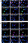
Localization of transiently expressed pp28 deletion mutants in the ERGIC. pp28 deletion mutants were transfected into Cos-7 cells. At day 2 posttransfection, the cells were fixed with 3% paraformaldehyde and examined by fluorescence microscopy as described in Materials and Methods. (A) Wild-type pp28EGFP (pp28WTEGFP) or pp28MutEGFP fluorescence is green, and organelle (p115, ERGIC marker) fluorescence is red (Texas Red). (B) Wild-type pp28EGFP (pp28WTEGFP) or pp28MutEGFP fluorescence is green, and organelle (calreticulin, ER marker) fluorescence is red (Texas Red). Nuclei are pseudocolored in blue following staining with Hoechst dye.
FIG. 3.
FIG. 3.
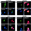
pp28 localization in the assembly compartment requires late gene expression. HF cells (3 × 10⁶ cells) were electroporated with approximately 5 μg of pp28EGFP and were either left uninfected (A) or infected with HCMV 48 h later at an MOI of 0.2 (B to D). Individual wells were treated with medium control (no drug, control) (B), ganciclovir (GCV) (C), or BDCRB (D). Cells were fixed 120 h postinfection, stained with anti-gM/gN and anti-IE-1 MAbs, and developed with TRITC anti-mouse IgG secondary antibodies. Nuclei were stained with Hoechst dye (blue) and IE-1 (red) in infected cells. The arrow in B demarcates the assembly compartment as indicated by gM/gN staining. Note that pp28EGFP was present in the assembly compartment of >90% of cells in control wells compared to <10% of cells in GCV-treated cultures.
FIG. 4.
FIG. 4.
Localization of pp28 deletion mutants to the assembly compartment late in infection in HCMV-infected HF cells. HF cells were electroporated with approximately 5 μg of expression plasmids encoding wild-type pp28EGFP (pp28WT-EGFP), pp28Mut40EGFP, pp28Mut50EGFP, pp28Mut61EGFP, pp28ΔacEGFP, or pp28ΔacCtTR-Myc and infected 2 days later with HCMV at an MOI of 0.2. The cells were harvested at day 7 postinfection, fixed with 3% paraformaldehyde, stained with anti-IE-1 MAbs combined with anti-GM130 MAbs (pp28EGFP, pp28Mut40EGFP, pp28Mut50EGFP, pp28Mut61EGFP, and pp28ΔacEGFP) to localize the AC to a secretory compartment or with anti-IE-1 MAbs only (pp28ΔacCtTR-Myc) followed by TRITC-labeled anti-mouse IgG to identify infected cells (red nuclei) and examined by confocal microscopy. The expression of the pp28ΔacCtTR-Myc-tagged mutant was detected with an anti-Myc MAb followed by FITC anti-mouse IgG (green fluorescence). The AC can be seen in the juxtanuclear position in cells expressing pp28EGFP, pp28Mut61EGFP, and pp28Mut50EGFP. Note the weak staining from the GM130 MAb staining of the Golgi compartment that is surrounding the AC in cells expressing wild-type pp28EGFP, pp28Mut40EGFP, pp28Mut50EGFP, and pp28Mut61EGFP. The GM130 reactivity in infected cells expressing pp28ΔacEGFP cannot be appreciated in this photograph.
FIG. 5.
FIG. 5.
Altered morphology of the secretory compartment following HCMV infection. HF cells were electroporated with approximately 5 μg of plasmids encoding ERGIC53 (ERGIC marker), mannosidase II (Golgi marker), and Gal T (TGN marker) proteins fused to EGFP and were either (A) left uninfected or (B) infected with HCMV at an MOI of 0.2 36 h after electroporation. The infected cells were incubated for 7 days prior to fixation as described in Materials and Methods. Cells that were fixed 7 days postinfection were reacted with anti-pp28 MAb followed by TRITC anti-mouse IgG to show the assembly compartment. Nuclei were stained (blue) with Hoechst dye. The arrow demarcates the assembly compartment.
FIG. 6.
FIG. 6.
Iodixanol density gradient fractionation of HCMV-infected HF cells transfected with pp28 mutants. HF cells were electroporated with approximately 5 μg of the designated pp28MutEGFP expression plasmids and infected 2 days later with HCMV at an MOI of 0.2. The cells were harvested at day 6 postinfection and fractionated by centrifugation through iodixanol (Optiprep; Sigma Co., St. Louis, Mo.) gradients as described in Materials and Methods. The gradient was fractionated by removing 1-ml fractions from the top; thus, fraction 1 represents the top of the gradient, and fraction 10 represents the bottom of the gradient. (A) Gradient fractions were analyzed by Western blot. Proteins were detected with specific antibodies, anti-pp28 MAb for WT pp28 and pp28Mut40EGFP, pp28Mut50EGFP, and pp28ΔacEGFP and developed with 125I-protein A. MAbs reactive with cellular proteins specific for compartments of the secretory pathway, anti-GM130 and anti-CD63, were used to localize pp28 and pp28 mutants to different intracellular compartments. (B) Results of panel A were analyzed by densitometry [density (%) = peak density of each fraction/total density of signal from protein in all gradient fractions × 100]. The top of B is a graphic comparison of the pattern of fractionation of virus-encoded pp28 to those of pp28Mut40EGFP, pp28Mut50EGFP, and pp28ΔacEGFP. Viral pp28 (WT) (▪), pp28Mut40EGFP (□), pp28Mut50EGFP (•), and pp28ΔacEGFP (○) are depicted. The bottom of B represents a comparison of the pattern of fractionation from viral pp28 to those of subcellular organelles (GM130, Golgi; CD63, late endosome). Viral pp28 (WT) (▪), GM130 (▴), and CD63 (▵) are depicted.
FIG. 7.
FIG. 7.
Construction of UL99 recombinant BACs containing mutations in pp28. (A) Recombinant BACs containing pp28 deletion mutants were generated by the insertion of single nucleotide (nt) changes in the UL99 coding sequence that altered wild-type codons to translational stops. Mutations were confirmed by sequence analysis. Revertant or repaired BACs were made by oligonucleotide-directed repair of the single nucleotide mutations. Recombinant viruses were recovered by electroporation of BAC DNA into HF cells. (B) Successful recovery of infectious viruses is listed on the right. In all cases, at least three independent attempts were made to recover infectious virus. The acidic cluster (ac) deletion BAC was made by the deletion of nucleotides 130 to 177 in the UL99 coding sequence. A virus was recovered from this pp28Δac BAC, but it formed small and very slowly expanding plaques. We could not isolate cell-free infectious virus from these cultures, nor did the plaques expand through the monolayer.
FIG. 8.
FIG. 8.
Expression of truncated pp28 in HF cells infected with UL99 recombinant virus. HF cells were infected with recombinant virus at an MOI of 0.1. The cells were harvested at day 5 postinfection, lysed, and detected with anti-pp28 MAb by Western blot analysis as described in Materials and Methods. Arrows indicate the mass predicted (kilodaltons) from the amino acid sequence of truncated pp28. An asterisk (*) indicates the migration of wild-type pp28.
FIG. 9.
FIG. 9.
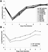
Replication kinetics of wild-type and recombinant viruses. Virus titer was quantified by a fluorescence-based virus infectivity assay on HF cells infected with WT or recombinant viruses at the indicated times after an initial infection using an MOI of 0.1. Results are expressed as log10 copies/milliliter (A) wild-type (▪) versus STOP61 (•), STOP90 (▴), STOP123 (⧫), STOP155 (*), Rev33 (+), Rev61 (○), Rev90 (▵), Rev123 (⋄), or Rev155 (×) virus. (B) Wild-type (▪) versus STOP50 (□) virus.

Similar articles

Cited by

References

    1. Abate, D. A., S. Watanabe, and E. S. Mocarski. 2005. Major human cytomegalovirus structural protein pp65 (ppUL83) prevents interferon response factor 3 activation in the interferon response. J. Virol. 78:10995-11006. - PMC - PubMed
    1. Andreoni, M., M. Faircloth, L. Vugler, and W. J. Britt. 1989. A rapid microneutralization assay for the measurement of neutralizing antibody reactive with human cytomegalovirus. J. Virol. Methods 23:157-167. - PubMed
    1. Appenzeller, C., H. Andersson, F. Kappeler, and H. P. Hauri. 1999. The lectin ERGIC-53 is a cargo transport receptor for glycoproteins. Nat. Cell Biol. 1:330-334. - PubMed
    1. Billstrom, M. A., and W. J. Britt. 1995. Postoligomerization folding of human cytomegalovirus glycoprotein B: identification of folding intermediates and importance of disulfide bonding. J. Virol. 69:7015-7022. - PMC - PubMed
    1. Bonifacino, J. S., and L. M. Traub. 2003. Signals for sorting of transmembrane proteins to endosomes and lysosomes. Annu. Rev. Biochem. 72:395-447. - PubMed

Publication types

LinkOut - more resources