. 2006 Jul;3(7):e263.
doi: 10.1371/journal.pmed.0030263. Epub 2006 May 23.

Genome microevolution of chikungunya viruses causing the Indian Ocean outbreak

Affiliations

Genome microevolution of chikungunya viruses causing the Indian Ocean outbreak

Isabelle Schuffenecker et al. PLoS Med. 2006 Jul.

Abstract

Background: A chikungunya virus outbreak of unprecedented magnitude is currently ongoing in Indian Ocean territories. In Réunion Island, this alphavirus has already infected about one-third of the human population. The main clinical symptom of the disease is a painful and invalidating poly-arthralgia. Besides the arthralgic form, 123 patients with a confirmed chikungunya infection have developed severe clinical signs, i.e., neurological signs or fulminant hepatitis.

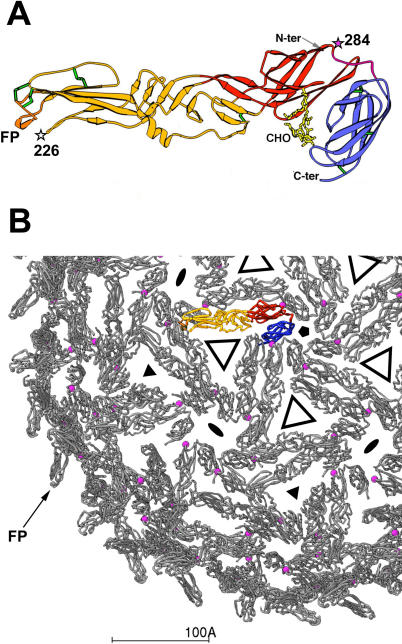
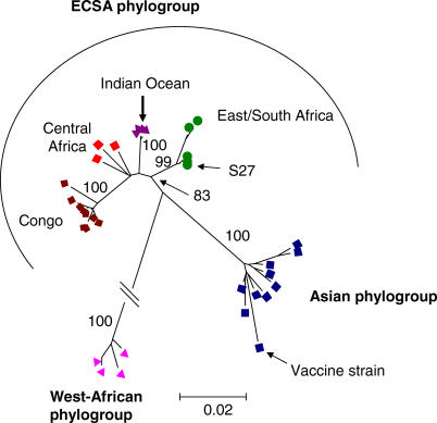
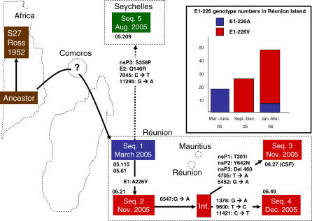
Methods and findings: We report the nearly complete genome sequence of six selected viral isolates (isolated from five sera and one cerebrospinal fluid), along with partial sequences of glycoprotein E1 from a total of 127 patients from Réunion, Seychelles, Mauritius, Madagascar, and Mayotte islands. Our results indicate that the outbreak was initiated by a strain related to East-African isolates, from which viral variants have evolved following a traceable microevolution history. Unique molecular features of the outbreak isolates were identified. Notably, in the region coding for the non-structural proteins, ten amino acid changes were found, four of which were located in alphavirus-conserved positions of nsP2 (which contains helicase, protease, and RNA triphosphatase activities) and of the polymerase nsP4. The sole isolate obtained from the cerebrospinal fluid showed unique changes in nsP1 (T301I), nsP2 (Y642N), and nsP3 (E460 deletion), not obtained from isolates from sera. In the structural proteins region, two noteworthy changes (A226V and D284E) were observed in the membrane fusion glycoprotein E1. Homology 3D modelling allowed mapping of these two changes to regions that are important for membrane fusion and virion assembly. Change E1-A226V was absent in the initial strains but was observed in >90% of subsequent viral sequences from Réunion, denoting evolutionary success possibly due to adaptation to the mosquito vector.

Conclusions: The unique molecular features of the analyzed Indian Ocean isolates of chikungunya virus demonstrate their high evolutionary potential and suggest possible clues for understanding the atypical magnitude and virulence of this outbreak.


Conflict of interest statement

Competing Interests: The authors have declared that no competing interests exist.

Figures

Figure 1
Figure 1. Localization of the E1 Changes on the 3D Structure Modelled from the Crystal Structure of SFV E1
(A) Ribbon diagram of E1, with domain I coloured red, domain II yellow, and domain III blue. Green tubes mark the disulfide bonds. The fusion peptide, at the tip of the molecule (in domain II) is coloured orange and labelled. The N-terminus and the C-terminus observed in the crystal (which is 30 aa upstream of the trans-membrane region) are also labelled. The two unique changes observed in the Indian Ocean isolates are indicated by stars and labelled: positions 226 (white) and 284 (magenta). (B) Partial representation (one octant, slightly extended) of the icosahedral E1 scaffold at the surface of the virion, viewed down a 5-fold symmetry axis. One E1 protomer is highlighted in colours, as in (A); all the others are represented in grey. The location of the some of the icosahedral symmetry axes are drawn as solid black symbols: pentagon for 5-fold axis, triangle for 3-fold axes, ellipse for 2-fold axes (which in the T = 4 lattice of alphaviruses are coincident with quasi 6-fold axes). Open triangles indicate roughly the location of the E2 trimers that interact tightly with E1, covering domain II and the fusion peptide, and presenting the main antigenic sites. The open triangles mark also quasi 3-fold symmetry axes of the T = 4 surface icosahedral lattice. A magenta ball marks the location of Glu 284, at an inter-E1 protomer contact site. This contact is propagated 240 times at the surface lattice (note all pink balls drawn on the grey protomers). Note that the fusion peptide, in orange, is pointing up and away from contacts with other E1 protomers. This is more easily seen at the periphery of the virion, where one of them is labelled (FP). In the virion, this region of E1 is not accessible, covered underneath the E2 molecule [ 20].
Figure 2
Figure 2. Phylogenetic Relationships among Chikungunya Isolates Based on Partial E1 Nucleotide Sequences
Isolates from the Indian Ocean outbreak (Réunion, Seychelles, Mayotte, Mauritius, and Madagascar) represent a distinct clade within a large East-, Central-, and South-African (ECSA) phylogroup. Bootstrap resampling values are indicated at major nodes. The branch leading to West-African phylogroup (of length of approximately 15%) was shortened for convenience. Maximum-likelihood analysis resulted in a very similar phylogeny.
Figure 3
Figure 3. Proposed Evolutionary Scenario of CHIKV Isolates from the Indian Ocean Outbreak
The scenario is based on six genome sequences determined by direct sequencing of RT-PCR products obtained using RNA extracts as templates; the sequences (Seq.) thus correspond to consensus sequences of the possible mixture of coexisting genomes (quasispecies). Inset: number of cases of E1-226A and E1-226V at different time intervals in Réunion Island, based on partial E1 sequences. E1-226V was observed in consensus sequences 2, 3, and 4, and therefore most E1-226V isolates genotyped based on partial E1 sequences are likely related to these genotypes. However, the independent appearance of E1-226V in other genotypes cannot be excluded. The location, size, and relative position of the islands and the African border are indicative. Consensus sequence 1 was obtained from a Réunion patient who travelled back from Comoros in March 2005, and from a Réunion Island patient. Sequences 2–4 were sampled in Réunion Island; sequence 5 was sampled in the Seychelles. The strain codes are given under the corresponding sequences. The question mark in Comoros Archipelago indicates that Sequence 1 possibly originates from Comoros. The dotted link between Sequence 1 and Sequence 5 indicates that the transmission route is hypothetical. CSF, cerebrospinal fluid; Int., deduced intermediate sequence, which was not sampled.

References

    1. Strauss EG, Strauss JH. Structure and replication of the alphavirus genome. In: Schlesinger S, Schlesinger MJ, editors. The Togaviridae and Flaviviridae. New York: Plenum Press; 1986. pp. 35–90.
    1. Porterfield JH. Antigenic characteristics and classification of the Togaviridae . In: Schlesinger R, editor. The Togaviruses. New York: Academic Press; 1980. pp. 13–46.
    1. Ross RW. The Newala epidemic. III. The virus: Isolation, pathogenic properties and relationship to the epidemic. J Hyg. 1956;54:177–191. - PMC - PubMed
    1. Jupp PG, McIntosh BM. Chikungunya disease. In: Monath TP, editor. The Arboviruses: Epidemiology and ecology. Boca Raton (Florida): CRC Press; 1988. pp. 137–157.
    1. Johnston RE, Peters CJ. Alphaviruses associated primarily with fever and polyarthritis. In: Fields BN, Knipe DM, Howley PM, editors. Fields virology. Philadelphia: Lippincott-Raven Publishers; 1996. pp. 843–898.

Publication types