. 2006 May 22;173(4):559-70.
doi: 10.1083/jcb.200601014. Epub 2006 May 15.

M line-deficient titin causes cardiac lethality through impaired maturation of the sarcomere

Affiliations

M line-deficient titin causes cardiac lethality through impaired maturation of the sarcomere

Stefanie Weinert et al. J Cell Biol. .

Abstract

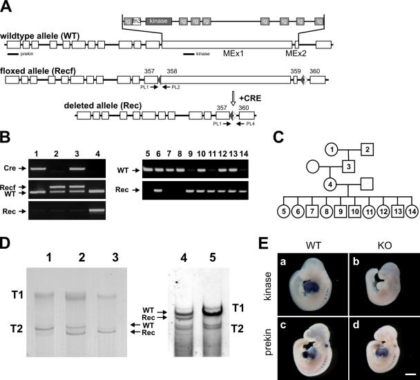
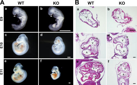
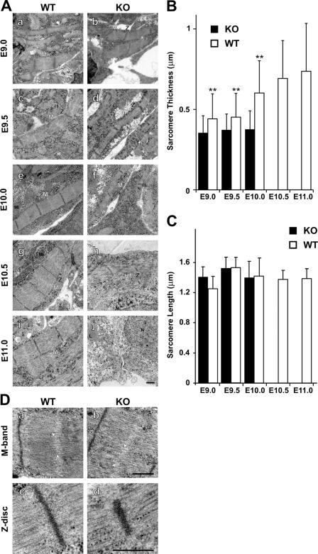
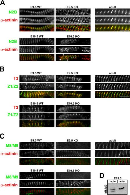
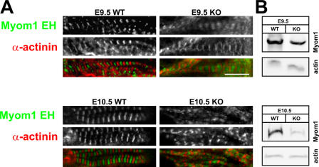
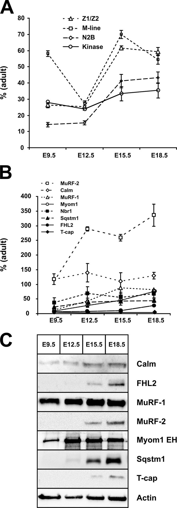
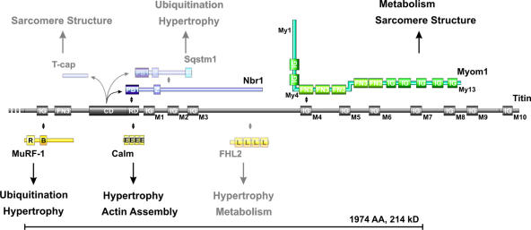
Titin, the largest protein known to date, has been linked to sarcomere assembly and function through its elastic adaptor and signaling domains. Titin's M-line region contains a unique kinase domain that has been proposed to regulate sarcomere assembly via its substrate titin cap (T-cap). In this study, we use a titin M line-deficient mouse to show that the initial assembly of the sarcomere does not depend on titin's M-line region or the phosphorylation of T-cap by the titin kinase. Rather, titin's M-line region is required to form a continuous titin filament and to provide mechanical stability of the embryonic sarcomere. Even without titin integrating into the M band, sarcomeres show proper spacing and alignment of Z discs and M bands but fail to grow laterally and ultimately disassemble. The comparison of disassembly in the developing and mature knockout sarcomere suggests diverse functions for titin's M line in embryonic development and the adult heart that not only involve the differential expression of titin isoforms but also of titin-binding proteins.


Figures

Figure 1.
Figure 1.
Conversion of the inducible into a constitutive titin kinase region knockout. (A) Outline of the exon/intron structure of titin's M-line region and location of the genotyping primers (PL1, 2, and 4) and in situ probes (prekin and kinase). Cre-mediated recombination leads to the deletion of exons 358 and 359 (MEx1 and 2). (B) PCR-based genotyping of the protamine-Cre transgenic mouse (lane 1) and Recf mouse with loxP sites flanking MEx1 and 2 (lane 2) that were mated to obtain double heterozygotes (lane 3). After germline recombination, offspring contain the deleted Rec allele (lane 4). PCR analysis of embryos at E9.5 confirms early embryonic survival of homozygous knockouts (lanes 9, 11, and 14). (C) Pedigree for animals analyzed in B. (D) SDS-agarose gels of wild-type (lane 1), heterozygous (lane 2), and knockout hearts (lane 3) of E9.5 animals and adult heterozygous (lane 4) and wild-type animals (lane 5). Titin proteins of the predicted sizes are expressed in knockout animals. Because embryonic titin is expressed as a larger isoform, differences in migration are more prevalent in T2 titin. Truncated titin (Rec) is more stable in the embryo compared with adult heterozygotes (ratio of wild-type/knockout protein in lanes 2 and 4). Homozygous adults could not be obtained. (E) In situ hybridization of whole-mount E9.5 embryos using an antisense probe directed against the kinase region (a and b) and a probe that recognizes a region upstream of the kinase domain (prekin in c and d). Both heart and somites are stained in all controls, whereas the kinase probe in knockout animals does not produce a signal (b). Note the smaller body size of titin M-line knockout animals (KO). WT, wild type. Bar, 500 μm.
Figure 2.
Figure 2.
Knockout embryos develop normally up to E9.0, followed by a failure to thrive. (A) The wild-type (WT) and knockout (KO) embryos at E9.0 (a and b) develop normally with appropriate cardiac size and function. The mutant embryo at E10.0 (d) appears normal, including normal cardiac morphology, but is small for its age. At E11.0, embryonic and cardiac development of the mutant is delayed with reduced body size and pericardial hemorrhage (f). Arrows point to the heart. (B) Histological analysis demonstrates normal development of the left ventricle and myocardium until E9.0. At E10.0, proper looping takes place, but ventricular wall thickness and trabeculation are reduced compared with wild-type animals (f vs. e; and Fig S2, available at http://www.jcb.org/cgi/content/full/jcb.200601014/DC1). At E11.0, the difference in wall thickness becomes more prominent and is accompanied by cellular infiltration of the knockout pericardium (f, arrowheads). A, atrium; LA, left atrium; RA, right atrium; V, ventricle; M, myocardium; T, trabeculation. Bars, 500 μm.
Figure 3.
Figure 3.
Ultrastructural analysis of cardiac sarcomere maturation. (A) Sarcomeres assemble in both wild-type (WT) and knockout animals (KO), but lateral growth is impaired from day 10 in knockout animals followed by sarcomere disassembly (h and j). At E11, nearly all sarcomeric structures are dissolved. (B) Myofibril diameter was quantified for >50 fibers in duplicate with two animals per group. The lateral growth of knockout myofibrils lags behind the increase in wild-type myofibril diameter. The loss of sarcomere structure precludes the analysis of E10.5 and 11.0 sarcomeres in knockout animals. **, P < 0.01. (C) Sarcomere length was not changed in wild-type versus knockout animals before disassembly at E10.5. Error bars represent SD. (D) M-band and Z-disc structures at E10.0 do not differ significantly between knockouts and wild-type animals, and there is no change in the A/I alignment. Slight variation in M-band alignment is present in both knockout and wild-type animals (arrowheads in a and b). Embryonic sarcomeres do not display an electron-dense M band. L, lipid; Mi, mitochondrium; N, nucleus; Z, Z disc; M, M band. Bars, 0.5 μm.
Figure 4.
Figure 4.
Integration of titin into the sarcomere. Coimmunostaining with antibodies directed against α-actinin and titin's N2B region (A), the A/I region (B), and the M-line region (C) reveals that titin is expressed and incorporated into the I band of the sarcomere in both wild-type (WT) and knockout (KO) animals (A). The N2B region is located proximal to the Z disc, and, thus, N2B staining partially overlaps with α-actinin staining at E9.5 (yellow). In knockouts at day 10.5, sarcomeres disassemble with few areas of striation remaining (open arrowheads). In areas of disassembly where α-actinin localizes in spotted aggregates (closed arrowheads), the N2B region is distributed diffusely. (B) The T3 antibody stains titin at the beginning of the A band. Correct localization in wild-type and knockouts (doublets marked with arrowheads) indicates that titin transitions properly into the A band. (C) Unlike the N2B region and the T3 epitope, titin's M line (M8/M9) is not integrated into the sarcomere in the absence of the kinase region. Although wild-type sarcomeres at different stages of embryonic development and in the adult animal show the expected striated pattern with alternating Z-disc and M-band epitopes, knockout animals fail to incorporate titin into the M band. Already at E9.5, titin's COOH terminus localizes diffusely. (D) To confirm the presence of titin's M8/M9 epitope in the kinase region–deficient protein, we used Western blot analysis of the wild-type and truncated titin in heterozygous embryos. Bars, 5 μm.
Figure 5.
Figure 5.
M-band assembly in the absence of titin's M-line region. (A) Although titin is not incorporated into the M band, myomesin (Myom1 EH) localizes between Z discs (α-actinin staining). (B) The more diffuse myomesin staining in titin kinase region knockout (KO) animals is not related to the overexpression of myomesin, as demonstrated by Western blotting at both E9.5 and 10.5. Expression was normalized to actin. WT, wild type. Bar, 5 μm.
Figure 6.
Figure 6.
Sarcomere metabolism in embryonic development versus adulthood. Real-time RT-PCR analysis of titin (A) and its M line–binding proteins (B) demonstrates that transcript levels change by up to two orders of magnitude from early cardiac development to adulthood. (A) The cardiac-specific N2B region is barely expressed in the embryonic heart. Differences in Z-disc and M-line levels indicate differences in the ratio of full-length titin to the NH2-terminal novex isoforms, with more full-length titin expressed in the developing embryo, as depicted in Fig. S2 A (available at http://www.jcb.org/cgi/content/full/jcb.200601014/DC1). (B) Except for calmodulin, MuRF-2, and Nbr1, transcript levels of most binding proteins are <20% of adult levels at E9.5. The proposed kinase substrate T-cap shows the lowest embryonic expression of all transcripts tested. (C) Most M-line titin–binding proteins can be detected by Western blotting only after E9.5, when cardiac pathology in knockout animals is already present. Notable exceptions are MuRF-1, which is expressed at similar levels throughout embryonic development, calmodulin, and myomesin. Error bars represent SD. Calm, calmodulin; FHL2, four and a half LIM-only protein 2; Nbr1, neighbor of BRCA1 gene 1; Sqstm1, sequestosome 1; MuRF, muscle-specific RING finger protein; T-cap, titin cap.
Figure 7.
Figure 7.
Signaling and structural functions of titin's M-line region. The titin M-line region (gray) mainly consists of Ig domains labeled M1–M10, a fibronectin (FN3), and a kinase domain (CD + RD). The 214-kD region deleted in our titin M line–deficient animals (bracket) contains binding sites for MuRF-1, Nbr1, calmodulin, FHL2 (signaling proteins in yellow), and myomesin (green). Myomesin integrates into the M band through interaction with myosin (My1), titin (My4), and dimerization (My13). In vitro substrates for the truncated soluble kinase are T-cap, p62, and Nbr1 (blue). The titin M-line region has been proposed to cover structural (sarcomere assembly) as well as signaling functions (hypertrophy/atrophy) through its multiple binding partners. Proteins expressed at low levels or below the detection limit of our Western blot in the embryonic heart are semitransparent. Protein–protein interactions are indicated by double-pointed arrows. The protein domains depicted are PB1 (Phox and Bem1p domain), Z (zinc-binding domain), U (ubiquitin-associated domain), IG (Ig-like domain), FN3 (fibronectin type 3 domain), CD (titin kinase catalytic domain), RD (titin kinase regulatory domain), R (RING finger), B (B box–type zinc finger), E (EF hand calcium-binding motif), and L (LIM domain).

References

    1. Agarkova, I., and J.C. Perriard. 2005. The M-band: an elastic web that crosslinks thick filaments in the center of the sarcomere. Trends Cell Biol. 15:477–485. - PubMed
    1. Agarkova, I., E. Ehler, S. Lange, R. Schoenauer, and J.C. Perriard. 2003. M-band: a safeguard for sarcomere stability? J. Muscle Res. Cell Motil. 24:191–203. - PubMed
    1. Ahuja, P., E. Perriard, J.C. Perriard, and E. Ehler. 2004. Sequential myofibrillar breakdown accompanies mitotic division of mammalian cardiomyocytes. J. Cell Sci. 117:3295–3306. - PubMed
    1. Auerbach, D., B. Rothen-Ruthishauser, S. Bantle, M. Leu, E. Ehler, D. Helfman, and J.C. Perriard. 1997. Molecular mechanisms of myofibril assembly in heart. Cell Struct. Funct. 22:139–146. - PubMed
    1. Auerbach, D., S. Bantle, S. Keller, V. Hinderling, M. Leu, E. Ehler, and J.C. Perriard. 1999. Different domains of the M-band protein myomesin are involved in myosin binding and M-band targeting. Mol. Biol. Cell. 10:1297–1308. - PMC - PubMed

Publication types

MeSH terms