Comparative Study
. 2006 Jun;127(6):749-54.
doi: 10.1085/jgp.200609527. Epub 2006 May 15.

Ca2+-activated Cl- current from human bestrophin-4 in excised membrane patches

Affiliations
Comparative Study

Ca2+-activated Cl- current from human bestrophin-4 in excised membrane patches

Takashi Tsunenari et al. J Gen Physiol. 2006 Jun.

Abstract

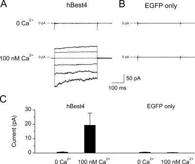
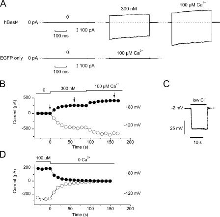
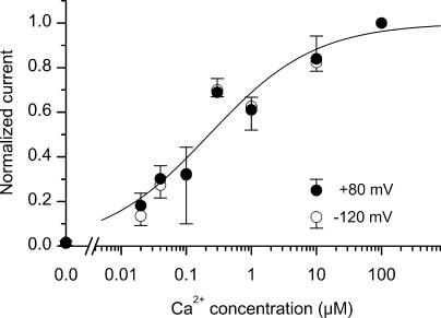
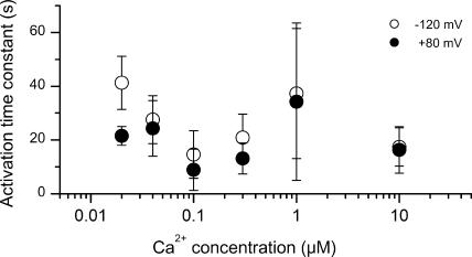
Bestrophins are a newly discovered family of Cl(-) channels, some members of which are activated by intracellular Ca(2+). So far, all studies were carried out with whole-cell recordings from plasmid-transfected cultured cells, so it is unclear whether Ca(2+) activates bestrophin through a metabolic mechanism or in a more direct way. We report here experiments that addressed this question with excised, inside-out membrane patches. We chose human bestrophin-4 (hBest4) for heterologous expression because it gave particularly large Cl(-) currents when expressed, thus allowing detection even in excised membrane patches. hBest4 gave a negligible Cl(-) current in a Ca(2+)-free solution on the cytoplasmic (bath) side, but produced a Cl(-) current that was activated by Ca(2+) in a dose-dependent manner, with a K(1/2) of 230 nM. Thus, Ca(2+) appears to activate the bestrophin Cl(-) channel without going through a freely diffusible messenger or through protein phosphorylation. Because the activation and deactivation kinetics were very slow, however, we cannot exclude the involvement of a membrane-associated messenger.


Figures

Figure 1.
Figure 1.
Cl current of hBest4 in inside-out membrane patches excised from transfected CHO-K1 cells. (A) Cell cotransfected with hBest4 plasmid and EGFP plasmid. (B) Cell transfected with EGFP plasmid alone (negative control). In each case, the same patch was alternatively exposed to near-zero Ca2+ and 100-nM Ca2+ in the bath. The current traces were produced by 350-ms voltage steps from a holding potential of 0 mV to voltages between −120 and +80 mV in 40-mV increments. Both of the patch pipette solution and the bath solution contained NMDG+ and Cl as major ions. (C) Collected results of experiments shown in A and B, measured at the end of the 350-ms voltage to +80 mV (mean ± SEM). Left, nine patches; right; seven patches.
Figure 2.
Figure 2.
Ca2+ dependence of hBest4 Cl current. (A) Top, currents recorded from an excised patch of hBest4-transfected HEK293 cell at free Ca2+ concentrations of near-zero, 300 nM, and 100 μM. In each panel, the membrane voltage was stepped from 0 to −120 and +80 mV for 350 ms. Bottom, control experiment on an excised patch from a HEK293 cell mock-transfected with EGFP plasmid alone. (B) Complete recordings of the same patch as shown in the top of A to indicate the time course of activation of hBest4 current by Ca2+. Each point was derived from a set of measurements as shown in the top of A, showing the current amplitude at the end of the 350-ms pulse to −120 mV (open symbol) and +80 mV (closed symbol). The arrows indicate the time points at which the measurements in the top of A were obtained. The current activation time course at 300 nM Ca2+ in B could be described by a single exponential with time constant of 19 s at −120 mV and 10 s at +80 mV. (C) Solution-exchange time course measured with the same patch shown in A and B. The solution exchange was relatively fast. Membrane voltage was recorded in zero-current clamp mode. A high-Cl solution (135 mM Cl, E Cl = −2.7 mV) containing 100 μM Ca2+ was replaced by a low-Cl solution (23.6 mM Cl, E Cl = −47 mV) containing the same free [Ca2+]. No compensation for liquid-junction potential has been made in the trace. After compensation, the initial voltage would correspond to −1.5 mV, and reach −45 mV within 1–2 s after the onset of the solution exchange. (D) Same kind of experiment as in B, but from a different patch and showing the decline time course of the current.
Figure 3.
Figure 3.
Dependence of hBest4 Ca2+-activated Cl current on free Ca2+ concentration. Same procedure as in Fig. 2 B, including the order of solution application: near-zero Ca2+ followed by an intermediate Ca2+ concentration, and then by 100 μM Ca2+. The plotted currents have been normalized with respect to the current at 100 μM Ca2+. Filled circles represent mean values (±SD) at +80 mV, and open circles represent mean values at −120 mV (3–5 patches each). The smooth curve is the Hill equation with K1/2 = 230 nM and a Hill coefficient of 0.53.
Figure 4.
Figure 4.
Time constant of current activation at different Ca2+ concentrations. Same patches as in Fig. 3. The values are mean ± SD.

References

    1. Clapham, D.E. 2003. TRP channels as cellular sensors. Nature. 426:517–524. - PubMed
    1. Deutman, A.F. 1969. Electro-oculography in families with vitelliform dystrophy of the fovea. Detection of the carrier state. Arch. Ophthalmol. 81:305–316. - PubMed
    1. Fischmeister, R., and H.C. Hartzell. 2005. Volume sensitivity of the bestrophin family of chloride channels. J. Physiol. 562:477–491. - PMC - PubMed
    1. Francois, J., A. De Rouck, and D. Fernandez-Sasso. 1967. Electro-oculography in vitelliform degeneration of the macula. Arch. Ophthalmol. 77:726–733. - PubMed
    1. Gallemore, R.P., B.A. Hughes, and S.S. Miller. 1998. a. Light-induced responses of the retinal pigment epithelium. In The Retinal Pigment Epithelium. M.F. Marmor and T.J. Wolfensberger, editors. Oxford University Press, Oxford. 175–198.

Publication types