. 2006 May 15;20(10):1353-64.
doi: 10.1101/gad.1387406.

Keratin 17 modulates hair follicle cycling in a TNFalpha-dependent fashion

Affiliations

Keratin 17 modulates hair follicle cycling in a TNFalpha-dependent fashion

Xuemei Tong et al. Genes Dev. .

Abstract

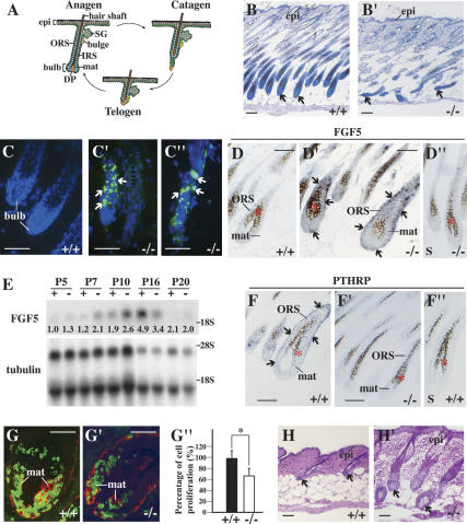
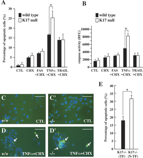
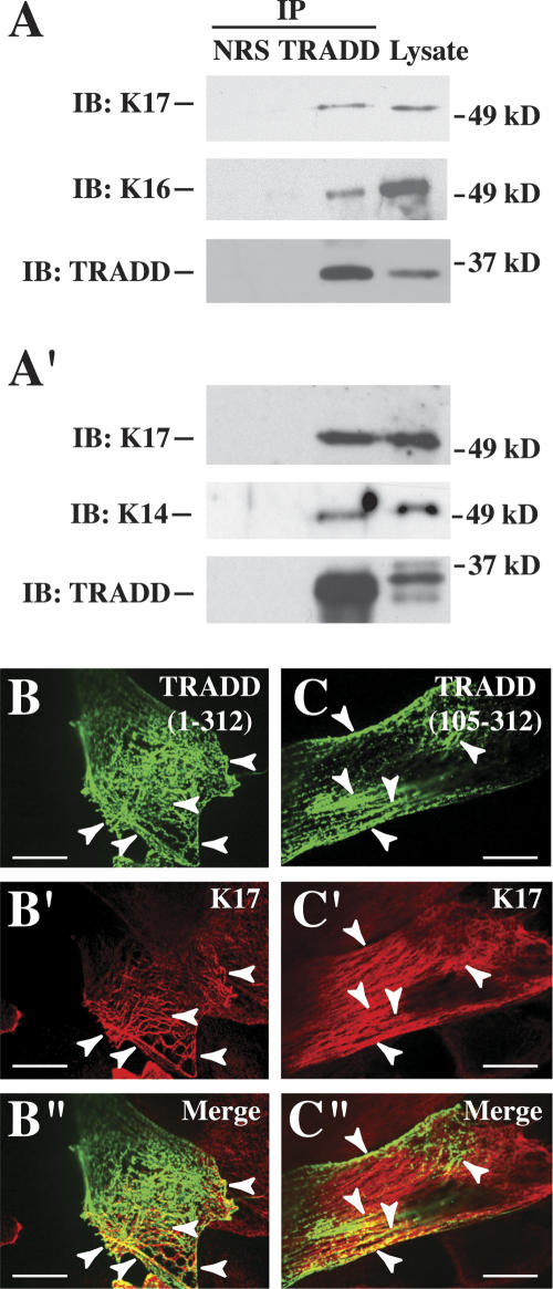
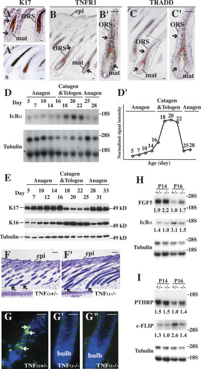
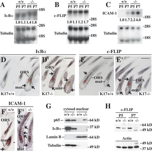
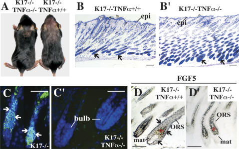
Mammalian hair follicles cycle between stages of rapid growth (anagen) and metabolic quiescence (telogen) throughout life. Transition from anagen to telogen involves an intermediate stage, catagen, consisting of a swift, apoptosis-driven involution of the lower half of the follicle. How catagen is coordinated, and spares the progenitor cells needed for anagen re-entry, is poorly understood. Keratin 17 (K17)-null mice develop alopecia in the first week post-birth, correlating with hair shaft fragility and untimely apoptosis in the hair bulb. Here we show that this abnormal apoptosis reflects premature entry into catagen. Of the proapoptotic challenges tested, K17-null skin keratinocytes in primary culture are selectively more sensitive to TNFalpha. K17 interacts with TNF receptor 1 (TNFR1)-associated death domain protein (TRADD), a death adaptor essential for TNFR1-dependent signal relay, suggesting a functional link between this keratin and TNFalpha signaling. The activity of NF-kappaB, a downstream target of TNFalpha, is increased in K17-null skin. We also find that TNFalpha is required for a timely anagen-catagen transition in mouse pelage follicles, and that its ablation partially rescues the hair cycling defect of K17-null mice. These findings identify K17 and TNFalpha as two novel and interdependent regulators of hair cycling.


Figures

Figure 1.
Figure 1.
Hair cycling defect in K17−/− skin. (A) Schematic of anagen, catagen, and telogen phases of the hair cycle. Adapted from Beaudoin et al. (2005) (© 2005 National Academy of Sciences). (B,B′) Histologic appearance of K17+/+ (wild-type; B) and K17−/− (B′) mouse backskin at P14. Shown are hematoxylin/eosin-stained longitudinal sections from backskin of age- and gender-matched mice. Arrows point to hair bulbs. Bars, 50 μm. (CC″) TUNEL (green) and Hoechst (blue) stainings of wild-type (C) and K17−/− (C′,C″) mouse skin at P14. Arrows point to TUNEL-positive nuclei. Bars, 50 μm. (DD′) In situ hybridization analysis of FGF5 mRNA in K17+/+ and K17−/− hair follicles at P7. Four percent paraformaldehyde-fixed frozen sections were hybridized with antisense (D,D′) or sense (D″) probes. Asterisks in DD″ denote melanin pigment. Arrows point to hybridization signals. Bars, 50 μm. (E) Northern blot analysis of RNA (20 μg) prepared from backskin of K17+/+ and K17−/− mice at P5, P7, P10, P16, and P20, and probed for FGF5 and β-tubulin mRNAs. Signal was quantitated in a PhosphorImager and normalized to β-tubulin. Average values of two independent experiments are reported below each lane. (FF″) In situ hybridization analysis of PTHRP mRNA in K17+/+ and K17−/− hair follicles at P12. Four percent paraformaldehyde-fixed frozen sections were hybridized with antisense (F,F′) or sense (F″) probes. Asterisks in FF″ denote melanin pigment. Arrows point to hybridization signals. Bars, 50 μm. (GG″) Dual immunostaining for BrdU (green) and keratin 14 (red) in hair bulbs from K17+/+ (G) and K17−/− (G′) mice at P5. Quantitation of BrdU-positive nuclei per surface area of bulb tissue is shown in G″. (*) P < 0.01. Bars, 20 μm. (H,H′) Histologic appearance of K17+/+ (H) and K17−/− (H′) mouse backskin at P22. Shown are hematoxylin/eosin-stained longitudinal sections from backskin of age- and gender-matched mice. Arrows point to dermal papillae (H) or hair bulbs (H′). Bars, 50 μm. (Bulb) Hair bulb; (DP) dermal papilla; (epi) epidermis; (IRS) inner root sheath; (mat) matrix; (ORS) outer root sheath; (SG) sebaceous gland.
Figure 2.
Figure 2.
K17-null keratinocytes show increased apoptosis following TNFα treatment. (A,B) Response of wild-type and K17−/− keratinocytes in primary culture to various proapoptotic treatment combinations. (CTL) DMSO-treated controls; (CHX) cycloheximide. Other treatments are self-explanatory. A shows the percentage of apoptotic cells based on TUNEL and Hoechst staining, while B shows a caspase activity assay (RFU, relative fluorescence units). Both A and B show that K17−/− keratinocytes are selectively more sensitive to TNFα + CHX treatment than wild type. (*) p < 0.01. (CD′) Representative micrographs from wild-type and K17−/− cultures treated with DMSO vehicle (CTL) or TNFα + CHX. Dual TUNEL (green) and Hoechst (blue) stainings are shown. Arrows denote TUNEL-positive nuclei. Bars, 20 μm. (E) Comparing the sensitivity of K17−/− keratinocytes (K17-null N-TF) to TNFα + CHX treatment after transfection with wild-type K17 cDNA (K17-null TF). Percentage of apoptotic cells is based on TUNEL and Hoechst stainings (see A). Cells re-expressing K17 are significantly less sensitive to treatment than K17−/− cells. P < 0.001.
Figure 3.
Figure 3.
Association between K17 and TRADD in skin keratinocytes. (A,A′) Immunoprecipitates from lysates prepared from wild-type mouse skin keratinocytes in primary culture, using normal rabbit serum (NRS) and two distinct anti-TRADD polyclonal antibodies, obtained from Santa Cruz Biotechnology (A) and Dr. M. Inagaki (Aichi Cancer Research Institute, Nagoya, Japan) (Inada et al. 2001) (A′). Precipitates were subjected to Western blotting with anti-K17, anti-K16, anti-K14, or anti-TRADD polyclonal antibody, as indicated at left. The migration of 37- and 49-kDa markers is shown at right. (BC″) Indirect immunofluorescence of wild-type keratinocytes in primary culture transiently transfected with EGFP-TRADD fusion contructs. (BB″) Full-length TRADD (312 residues long). (CC″) TRADD’s C terminus (residues 105–312). The signal detected (GFP, K17, or merge) is identified in the upper right corner. Arrowheads point to instances of colocalization. Bars, 30 μm.
Figure 4.
Figure 4.
Characterization of TNFα signaling in wild-type mouse skin. (AC′) In situ hybridization for K17 (A,A′), TNFR1 (B,B′), and TRADD (C,C′) mRNAs in hair follicles of P5–P7 wild-type mice. Four percent paraformaldehyde-fixed frozen sections were hybridized with antisense (A,B,C) or sense probes as controls (A′; data not shown) for the designated transcripts. The boxed areas in B and C are shown at higher magnification in B′ and C′, respectively. Asterisks denote melanin pigment. Arrows point to hybridization signals. (Bulb) Hair bulb; (epi) epidermis; (mat) matrix; (ORS) outer root sheath. Bars, 20 μm. (D,D′) Northern blot analysis of total RNA (20 μg) prepared from backskin of wild-type mice at different ages, and probed for IκBα and β-tubulin (loading control) mRNAs. (D′) Normalized signal intensity for IκBα mRNA, based on average values of two independent experiments. The P5 value, used as a reference, has been normalized to 1. Peak levels of IκBα mRNA (3.86 ± 0.02) occur at P20. (E) Western blot analysis of K17 and K16 levels in total protein extracts (2.5 μg) prepared from backskin of wild-type mice at different ages. The tubulin blot serves as a loading control. (F,F′) Histology of TNFα+/− (F) and TNFα−/− (F′) skin at P16. Shown are hematoxylin/eosin-stained staining longitudinal sections from backskin of age- and gender-matched mice. Arrows point to hair bulbs. Bars, 50 μm. (GG″) TUNEL (green) and Hoechst (blue) stainings of TNFα+/− (G) and TNFα−/− (G′,G″) skin tissue at P16. Bars, 50 μm. (H,I) Northern blot analysis of total RNA (20 μg) prepared from backskin of TNFα+/− or TNFα−/− at P14 and P16, and probed for FGF5 and IκBα (in H), PTHRP and c-FLIP (in I) mRNAs. Signal was quantitated by PhosphorImager and normalized to β-tubulin. Average values from two independent experiments are reported below each lane.
Figure 5.
Figure 5.
Increased NF-κB activity in K17-null skin. (A–C) Northern blot analysis of total RNA (20 μg) prepared from backskin of K17+/+ and K17−/− mice at P5 and P7, and probed for IκBα (A), c-FLIP (B), and ICAM-1 (C) mRNAs. Signal was quantitated by PhosphorImager and normalized to β-tubulin. The average values of two or three independent experiments are reported below each lane. (DF′) In situ hybridization analysis for IκBα (D,D′), c-FLIP (E,E′), and ICAM-1 (F,F′) mRNAs in backskin tissue from K17+/+ and K17−/− mice at P7. Sections were hybridized with antisense (DF′) or sense probes (as control; data not shown) for the designated transcripts. Asterisks denote melanin pigment. Arrows point to hybridization signals. Bars, 50 μm. (G) Western blot analysis of cytosol and nuclear fraction prepared from backskin of K17+/+ and K17−/− mice at P5 using anti-p65, anti-IκBα, anti-lamin B, or anti-tubulin antibody. Lamin B and tubulin serve as controls for both loading and fractionation. Note that there is more relA (p65) in the nuclear fraction and less IκBα protein in the cytosol fraction of K17−/− than K17+/+ control. (H) Western blot analysis of total proteins (30 μg) extracted from backskin of K17+/+ and K17−/− mice at P5 and P7 using an anti-c-FLIP antibody. Actin serves as a loading control.
Figure 6.
Figure 6.
Genetic interaction between K17 and TNFα pathway. (A, left) Rescue of hair loss in K17−/− TNFα−/− mice. A K17−/− littermate is shown at right for reference. (B,B′) Hematoxylin/eosin-stained skin tissue sections from K17−/− TNFα+/+ (B) and K17−/− TNFα−/− (B′) mice at P14. Arrows point to hair bulbs. Bars, 50 μm. (C,C′) TUNEL (green) and Hoechst (blue) dual stainings in backskin sections prepared from K17−/− TNFα+/+ (C) and K17−/− TNFα−/− (C′) mice at P14. Arrows point to TUNEL positive staining. Bars, 50 μm. (D,D′) In situ hybridization for FGF5 mRNA in K17−/− TNFα+/+ (D) and K17−/− TNFα−/− (D′) backskin at P7. Four percent paraformaldehyde-fixed frozen sections were hybridized with antisense (D,D′) or sense (not shown) probes. Bars, 50 μm. (Bulb) Hair bulb; (epi) epidermis; (mat) matrix; (ORS) outer root sheath. Asterisks denote melanin pigments in hair bulb.

References

    1. Basile J.R., Eichten A., Zacny V., Munger K. NF-κB-mediated induction of p21(Cip1/Waf1) by tumor necrosis factor α induces growth arrest and cytoprotection in normal human keratinocytes. Mol. Cancer Res. 2003;1:262–270. - PubMed
    1. Beaudoin G.M., III, Sisk J.M., Coulombe P.A., Thompson C.C. Hairless triggers reactivation of hair growth by promoting Wnt signaling. Proc. Natl. Acad. Sci. 2005;102:14653–14658. - PMC - PubMed
    1. Bender L.M., Morgan M.J., Thomas L.R., Liu Z.G., Thorburn A. The adaptor protein TRADD activates distinct mechanisms of apoptosis from the nucleus and the cytoplasm. Cell Death Differ. 2005;12:473–481. - PubMed
    1. Bernot K., McGowan K., Coulombe P.A. Keratin 16 expression defines a subset of epithelial cells during skin morphogenesis and the hair cycle. J. Invest. Dermatol. 2002;119:1137–1149. - PubMed
    1. Bernot K.M., Coulombe P.A., Wong P. Skin: An ideal model system to study keratin genes and proteins. Methods Cell Biol. 2004;78:453–487. - PubMed

Publication types

MeSH terms

Substances