. 2006 May 23;103(21):8233-8.
doi: 10.1073/pnas.0601992103. Epub 2006 May 15.

Fluoxetine targets early progenitor cells in the adult brain

Affiliations

Fluoxetine targets early progenitor cells in the adult brain

Juan M Encinas et al. Proc Natl Acad Sci U S A. .

Abstract

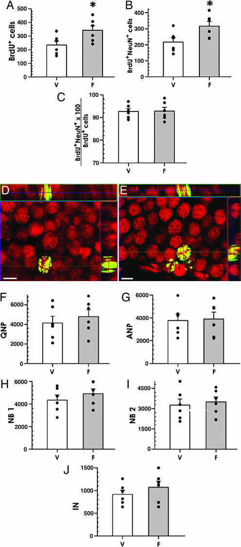
Chronic treatment with antidepressants increases neurogenesis in the adult hippocampus. This increase in the production of new neurons may be required for the behavioral effects of antidepressants. However, it is not known which class of cells within the neuronal differentiation cascade is targeted by the drugs. We have generated a reporter mouse line, which allows identification and classification of early neuronal progenitors. It also allows accurate quantitation of changes induced by neurogenic agents in these distinct subclasses of neuronal precursors. We use this line to demonstrate that the selective serotonin reuptake inhibitor antidepressant fluoxetine does not affect division of stem-like cells in the dentate gyrus but increases symmetric divisions of an early progenitor cell class. We further demonstrate that these cells are the sole class of neuronal progenitors targeted by fluoxetine in the adult brain and suggest that the fluoxetine-induced increase in new neurons arises as a result of the expansion of this cell class. This finding defines a cellular target for antidepressant drug therapies.


Conflict of interest statement

Conflict of interest statement: No conflicts declared.

Figures

Fig. 1.
Fig. 1.
Defining neuronal differentiation cascade in the DG. (A) Expression of endogenous nestin, detected by using a monoclonal antibody, in the DG of nestin-GFP transgenic mice; the pattern was the same in WT animals. (B) Expression of GFP in the DG of nestin-GFP transgenic mice; note that endogenous nestin in A is seen mostly in the processes, whereas GFP is present in the processes, the cytoplasm, and the nucleus. Tight packing of cells in the SGZ prevents accurate enumeration of nestin-GFP expressing cells. (C) Expression of CFPnuc, detected by using a polyclonal antibody in the SGZ of nestin-CFPnuc mice; transgene-expressing cells now are represented by their nuclei, thus making accurate cell counts even in densely packed areas possible. (DF) Expression of nestin (D) and GFP (E) in the SVZ of nestin-GFP mice and CFPnuc (F) in the SVZ of nestin-CFPnuc mice. Note that densely packed SVZ cells, which cannot be accurately counted in D or E, can be quantified easily in F. (G) GFP-expressing neural progenitor cells in the DG of the nestin-GFP mice. The soma of both QNP and ANP cells is seen in the SGZ; QNP cells carry vertical processes, which cross the granule cells layer and end as elaborated arbors in the molecular layer (processes also can be visualized by an antibody to GFAP). ANP cells lack the processes; during and immediately after division, they can be seen in close contact with QNP (note a QNP and an ANP cell above and beneath the dashed line). (HK) Asymmetric division of stem-like QNP cells in the DG generates ANP cells. (H) After BrdU labeling, cells with GFAP-labeled processes can be seen dividing (note the horizontal plane of division, dashed line) and generating daughter cells that are deposited below, do not carry processes or arbors, and do not stain for GFAP. (IK) Staining for GFAP (blue; I), BrdU (red; J), and CFPnuc (green; K). (L) A QNP cell (arrow) generates an ANP cell (arrowhead) through an asymmetric division, with the plane of division parallel to the SGZ. Nearby is a cluster of ANP cells (arrowheads) generated through symmetric divisions in the plane perpendicular to the SGZ. GFAP is red, and CFPnuc is green. (M and N) ANPs differentiate into NB1 cells, which start to express markers of young neurons. NB1 cells still are located in the SGZ, cease to express nestin or nestin-CFPnuc, and start to express PSA-NCAM (green), Dcx, and Prox-1. A small subclass of these cells (M) resembles the ANP cells morphologically and is the last cell population to incorporate BrdU. The majority of the NB1 cells (N) extends horizontal processes and does not incorporate BrdU. (O) NB1 cells evolve morphologically and are converted into NB2 cells. NB2 cells are largely confined to the SGZ and extend most of their processes horizontally; however, one process grows vertically or obliquely and extends into the GCL. These cells express PSA-NCAM (green), Dcx, Prox-1, and NeuN. (P) NB2 cells progress into IN. The morphology of these cells resembles that of mature granule neurons. They extend a single vertical apical process and have their soma in the granule cell layer. They express NeuN and Prox-1 but still express PSA-NCAM (green) and Dcx. (Scale bars: AF, 20 μm; G, H, LP, 5 μm.)
Fig. 2.
Fig. 2.
A schematic summary of the neuronal differentiation cascade in the DG. QNPs generate, through asymmetric divisions, the ANPs that, after several rounds of symmetric divisions, exit the cell cycle within 1–3 days and become postmitotic NB1 cells. Within next 15–21 days, NB1 cells mature into NB2 and then into IN with apical processes and basal axons and the soma located in the GCL. After an additional 10–15 days, INs acquire the characteristics of mature granule neurons, develop extensive branching, and send long axonal processes, forming the mossy fiber.
Fig. 3.
Fig. 3.
Fluoxetine increases cell proliferation in the adult DG. Chronic (15 days) fluoxetine treatment of adult (7 months) mice, analyzed 1 day after BrdU administration. Fluoxetine increases the number of BrdU-positive cells (A). (B and C) Representative photomicrographs of DG sections from animals treated with vehicle (B) and fluoxetine (C); dashed lines in B, C, E, and F outlines the external limits of the DG. Exposure to fluoxetine also increases the number of nestin-CFPnuc cells in the SGZ (D, histogram; E, section of the DG of a control animal; F, section of the DG of a fluoxetine-treated animal). Within total nestin-CFPnuc cells, the number of ANPs (H), but not QNPs (G), increases in response to fluoxetine. (Scale bars: 50 μm.) In all histograms, white bars correspond to the vehicle injections (V), and gray bars to the fluoxetine injections (F). Error bars show SEM. The results for individual animals (n = 8 per group in this figure) are shown as black dots. ∗, P < 0.05.
Fig. 4.
Fig. 4.
Fluoxetine increases NB1 cells in the adult DG. (A and B) Immunostaining for PSA-NCAM (green), and nestin-CFPnuc (red). Two cell types are distributed throughout the SGZ, often in close apposition to each other; however, they do not overlap, as illustrated in B (PSA-NCAM cell is red, and nestin-CFPnuc nuclei are green; note that colors are switched at low magnification for better visualization). (CG) Postmitotic precursors in the fluoxetine-treated DG of adult mice, analyzed 1 day after BrdU labeling. Fluoxetine increases the number of NB1 (C) but not of more advanced NB2 (D) or IN (E) cells. V, vehicle; F, fluoxetine. n = 8 per group. ∗∗, P < 0.01. F and G are representative photomicrographs of DG from control (injected with vehicle) (F) and fluoxetine-treated (G) animals. (Scale bars: A, 20 μm; B, 5 μm; F and G, 10 μm.)
Fig. 5.
Fig. 5.
Fluoxetine increases proliferation of ANP cells in the DG. (AD) Treatment with fluoxetine does not change the number of dividing (BrdU-labeled) QNPs (A) but increases division of ANPs (B). The fraction of BrdU-labeled QNP or ANP cells among total QNP or ANP cells, respectively, remains the same (C and D). V, vehicle; F, fluoxetine. n = 6 per group. ∗, P < 0.05. (EH) A cluster of BrdU-positive ANP cells between two QNPs in the DG of a fluoxetine-treated animal. QNP cells are identified by the presence of GFAP-positive processes. CFPnuc is shown in green (F), BrdU in red (G), and GFAP in blue (H). (Scale bar: 5 μm.)
Fig. 6.
Fig. 6.
Fluoxetine increases neurogenesis in the adult DG (30-day survival experiments). (AC) Chronic fluoxetine treatment of adult mice, analyzed 30 days after BrdU administration. Fluoxetine increases the number of BrdU-positive cells in the DG (A), and the number of BrdU and NeuN double-positive cells (B); the fraction of such cells among total BrdU-positive cells remains the same (C). (D and E) Representative photomicrographs of DG from control (vehicle) (D) and fluoxetine-treated (E) animals show that new cells became neurons, shown by immunostaining for BrdU (green) and NeuN (red). The orthogonal projections are shown to confirm double labeling throughout the extent of positive cells. (Scale bars: 10 μm.) (FJ) Fluoxetine treatment does not increase the number of neuronal progenitors when analyzed 30 days after the treatment. The histograms show the data for the QNP (F), ANP (G), NB1 (H), NB2 (I), and IN (J) cells. Changes did not reach the level of significance in none of the categories. V, vehicle; F, fluoxetine. n = 6 per group. ∗, P < 0.05.

References

    1. Wong M. L., Licinio J. Nat. Rev. Neurosci. 2001;2:343–351. - PubMed
    1. Whittington C. J., Kendall T., Fonagy P., Cottrell D., Cotgrove A., Boddington E. Lancet. 2004;363:1341–1345. - PubMed
    1. Ryan N. D. Lancet. 2005;366:933–940. - PubMed
    1. Malberg J. E., Eisch A. J., Nestler E. J., Duman R. S. J. Neurosci. 2000;20:9104–9110. - PMC - PubMed
    1. Jacobs B. L., Praag H., Gage F. H. Mol. Psychiatry. 2000;5:262–269. - PubMed

Publication types

Substances