. 2006 May 23;103(21):8209-14.
doi: 10.1073/pnas.0600919103. Epub 2006 May 15.

The positioning of cytoplasmic protein clusters in bacteria

Affiliations

The positioning of cytoplasmic protein clusters in bacteria

Stephen R Thompson et al. Proc Natl Acad Sci U S A. .

Abstract

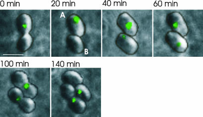
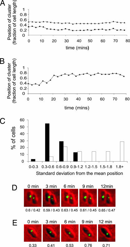
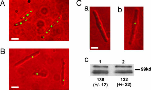
Cell division is a carefully orchestrated procedure. Bacterial cells have intricate mechanisms to ensure that genetic material is copied, proofread, and accurately partitioned into daughter cells. Partitioning now appears to also occur for some cytoplasmic proteins. Previously, using chromosomal fluorescent protein fusions, we demonstrated that a subset of Rhodobacter sphaeroides chemotaxis proteins colocalize to a discrete region within the bacterial cytoplasm. Using TlpT-yellow fluorescent protein as a marker for the position of the cytoplasmic protein clusters, we show most cells contain either one cluster localized at mid-cell or two clusters at the one-fourth and three-fourths positions of cell length. The number and positioning of these protein clusters depend on a previously unrecognized bacterial protein positioning factor, PpfA, which has homology to bacterial type I DNA partitioning factors. These data suggest that there is a mechanism involved in partitioning some cytoplasmic proteins upon cell division that is analogous to a mechanism seen for plasmid and chromosomal DNA.


Conflict of interest statement

Conflict of interest statement: No conflicts declared.

Figures

Fig. 1.
Fig. 1.
Positioning of the cytoplasmic protein clusters. Histograms show the distance of clusters from the farthest cell pole as a function of the cell length. Data are symmetrically distributed around mid-cell because the distance to the farthest cell pole was always measured. (A) Wild-type cells with one cluster found around mid-cell. (B) Wild-type cells with two clusters localized to the one-fourth and three-fourths positions. (C) ΔppfA cells never contained more than one cluster, which was randomly positioned within the cell.
Fig. 2.
Fig. 2.
Time-lapse images of the cytoplasmic protein cluster protein, TlpT-YFP. One cell containing two clusters divides into cells A and B. The single A cluster becomes two clusters early in cell growth, and each cluster migrates rapidly to positions at one-fourth and three-fourths of the cell length, where they remain until the cell divides. This positioning results in each daughter cell inheriting a cluster at about mid-cell. The B cell contains a single cluster throughout most of the cell cycle. This cluster is inherited by one of the daughter cells, and a new cluster is observed in the other daughter cell soon after septation. Contour lines showing the outlines of the cells are superimposed (the raw images are shown in Fig. 7). (Scale bar: 2 μm.)
Fig. 3.
Fig. 3.
Time-lapse images of the cytoplasmic protein cluster protein, TlpT-YFP, in cells deleted for the putative ATPase, PpfA. ΔppfA cells contain a maximum of one cytoplasmic cluster. After cell division, one daughter cell inherits the single cluster, and a new cluster becomes apparent in the other cell sometime later. Contour lines showing the outlines of the cells are superimposed (the raw images are shown in Fig. 8). (Scale bar: 2 μm.)
Fig. 4.
Fig. 4.
Short-interval time-course data on cytoplasmic cluster movement. Representative traces of the positions of cytoplasmic clusters of TlpT-YFP as a fraction of cell length in a wild-type background (A) and ΔppfA (B) are shown. The histogram (C) bins the results depending on the standard deviation from the mean position. Filled bars represent wild-type cells (n = 31), and open bars represent ΔppfA cells (n = 15). A low standard deviation of cluster position indicates a relatively fixed position within the cell. Clusters in the wild type are clearly less mobile than those in ΔppfA cells (Wilcoxon rank-sum test, P = 0.000133). Two example time courses are also shown in cells containing (D) and deleted for (E) ppfA. Underneath each cell is the position of each cluster as a fraction of cell length.
Fig. 5.
Fig. 5.
Fluorescence images of the cytoplasmic cluster position in filamentous cells. (A) Wild-type background. (B) ΔppfA. (C) Images have been treated in an identical manner and clearly show that the single cluster present in the ΔppfA strain (Cb) is brighter than any of the six clusters present in the wild-type background (only five clusters are visible in Ca, but six are observed with longer exposure times; data not shown). (Scale bars: 2 μm.) (Cc) Western blot analysis using anti-GFP antibody to crossreact with TlpT-YFP in filamentous cells containing (Cc1) and deleted for (Cc2) ppfA. Values represent the mean band intensity from six independent experiments (arbitrary units). The same samples were also probed with an antibody to CheA2, a component of the polar chemotaxis protein cluster whose level remained constant in both strains (data not shown).
Fig. 6.
Fig. 6.
The positioning of cytoplasmic chemotaxis protein clusters in the presence and the absence of PpfA. (A) Wild-type cells have a single cluster at around mid-cell. Before cell division, this cluster becomes two clusters that move to the one-fourth and three-fourths positions, resulting in each daughter cell inheriting a cluster at about mid-cell. When this happens late in the cell cycle, the second cluster may not be visible until after cell division. (B) In the absence of PpfA, cells never contain more than one cluster. This cluster appears mobile and increases in size as protein synthesis continues. Upon division, one daughter cell inherits the single cluster, and the other daughter cell synthesizes a new cluster de novo.

References

    1. Wadhams G. H., Armitage J. P. Nat. Rev. Mol. Cell Biol. 2004;5:1024–1037. - PubMed
    1. Bilwes A. M., Alex L. A., Crane B. R., Simon M. I. Cell. 1999;96:131–141. - PubMed
    1. Hess J. F., Oosawa K., Kaplan N., Simon M. I. Cell. 1988;53:79–87. - PubMed
    1. Levit M., Liu Y., Surette M., Stock J. J. Biol. Chem. 1996;271:32057–32063. - PubMed
    1. Alon U., Surette M. G., Barkai N., Leibler S. Nature. 1999;397:168–171. - PubMed

Publication types