. 2006 Jun 12;203(6):1447-58.
doi: 10.1084/jem.20060375. Epub 2006 May 15.

Vascular endothelial growth factor is an important determinant of sepsis morbidity and mortality

Affiliations

Vascular endothelial growth factor is an important determinant of sepsis morbidity and mortality

Kiichiro Yano et al. J Exp Med. .

Abstract

Sepsis, the systemic inflammatory response to infection, is a leading cause of morbidity and mortality. The mechanisms of sepsis pathophysiology remain obscure but are likely to involve a complex interplay between mediators of the inflammatory and coagulation pathways. An improved understanding of these mechanisms should provide an important foundation for developing novel therapies. In this study, we show that sepsis is associated with a time-dependent increase in circulating levels of vascular endothelial growth factor (VEGF) and placental growth factor (PlGF) in animal and human models of sepsis. Adenovirus-mediated overexpression of soluble Flt-1 (sFlt-1) in a mouse model of endotoxemia attenuated the rise in VEGF and PlGF levels and blocked the effect of endotoxemia on cardiac function, vascular permeability, and mortality. Similarly, in a cecal ligation puncture (CLP) model, adenovirus-sFlt-1 protected against cardiac dysfunction and mortality. When administered in a therapeutic regimen beginning 1 h after the onset of endotoxemia or CLP, sFlt peptide resulted in marked improvement in cardiac physiology and survival. Systemic administration of antibodies against the transmembrane receptor Flk-1 but not Flt-1 protected against sepsis mortality. Adenovirus-mediated overexpression of VEGF but not PlGF exacerbated the lipopolysaccharide-mediated toxic effects. Together, these data support a pathophysiological role for VEGF in mediating the sepsis phenotype.


Figures

Figure 1.
Figure 1.
Circulating levels of VEGF and PlGF in mouse and human models of sepsis. (a) The levels of IL-6 (plasma), TNF-α (serum), PlGF (plasma), and VEGF (plasma) in LPS-injected mice at the time points indicated; CTRL, control normal saline-injected mice. (b) Same as in a but in a CLP mouse model; sham, sham-operated control mice. (c) PlGF and VEGF levels in a mouse pneumonia model at 6 and 24 h. (d) PlGF and VEGF levels in a human endotoxemia model at the time points indicated. Subjects were administered LPS or saline (placebo). (e) Plasma PlGF and VEGF levels in randomly chosen patients with severe sepsis plotted against time in the intensive care unit (ICU). Each line represents an individual patient (P). Mouse data are expressed as means ± SD (a and c) or as means ± SEM (b) of three independent experiments. Human data from the endotoxemia model are expressed as means ± SEM. *, P < 0.05; **, P < 0.01; ***, P < 0.0001.
Figure 2.
Figure 2.
VEGF and PlGF protein levels in a mouse model of endotoxemia. Results of ELISA for VEGF (top) and PlGF (bottom) in tissues from mice injected with or without LPS at the time points indicated. *, P < 0.05; **, P < 0.01; ***, P < 0.0001. Error bars represent SD.
Figure 3.
Figure 3.
Effect of sFlt-1 overexpression on circulating levels of sFlt, VEGF, and PlGF in a mouse model of endotoxemia. Mice were injected with adenovirus overexpressing GFP (CTRL-ad) or sFlt-1 (sFlt-ad). 3 d later, the animals were administered saline (control) or LPS i.p. Blood samples were taken 24 h later and assayed for plasma levels of sFlt-1, free VEGF, and free PlGF. Data are expressed as means ± SD (error bars) of three independent experiments. *, P < 0.05; **, P < 0.01; ***, P < 0.0001.
Figure 4.
Figure 4.
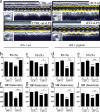
Effect of sFlt-1 overexpression on cardiac function in mouse models of sepsis. Mice were injected with adenovirus overexpressing GFP (CTRL-ad; C) or sFlt-1 (sFlt-ad; S) in a–c, f, and g. 3 d later, the animals were administered saline (control) or LPS i.p. or were subjected to CLP. Alternatively, mice were injected i.v. with PBS (C) or sFlt-1 peptide (S) 1 h after LPS injection or CLP procedure (d, e, h, and i). An echocardiogram and electrocardiogram were performed 24 h after saline or LPS injections or 26 h after CLP. (a) Representative M modes and two-dimensional images (insets) from an echocardiogram in an endotoxemia model. (b and d) Quantitative analysis of the fractional shortenings (FS) from an echocardiogram in an endotoxemia model. (f and h) Heart rate (HR) measurements on an electrocardiogram in an endotoxemia model. (c and e) Quantitative analysis of the fractional shortenings from an echocardiogram in a CLP model. (g and i) Heart rate measurements on an electrocardiogram in a CLP model. Analysis of variance was used for statistic analysis. Error bars represent SD. *, P < 0.05; **, P < 0.01; ***, P < 0.0001.
Figure 5.
Figure 5.
Effect of sFlt-1 overexpression on vascular permeability in a mouse model of endotoxemia. Mice were injected with adenovirus overexpressing GFP (CTRL-ad) or sFlt-1 (sFlt-ad). 3 d later, the animals were administered saline (control) or LPS i.p. 24 h later, the animals were injected i.v. with 0.1 ml of 1% Evans blue dye. After 40 min, the mice were perfused, and the brain (Br), lung (Lu), heart (H), liver (Li), kidney (Ki), and spleen (Sp) were harvested and incubated in formamide for 3 d to elute Evans blue dye. (left) Whole mount photomicrographs of organs. In each group, the left-most specimen is from control untreated mice, the middle specimen is from control adenovirus-treated endotoxemic mice, and the right specimen is from Ad–sFlt-1–treated endotoxemic mice (in the case of the kidney, two specimens are shown for each condition). (right) Quantitation of Evans blue extravasation (OD at 620 nm). Error bars represent SD. *, P < 0.05; **, P < 0.01; ***, P < 0.0001.
Figure 6.
Figure 6.
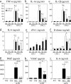
Effect of sFlt-1 overexpression on circulating cytokine levels and D-dimers in a mouse model of endotoxemia. (a–f) Mice were injected with adenovirus overexpressing GFP (CTRL-ad) or sFlt-1 (sFlt-ad). 3 d later, the animals were administered saline (control) or LPS i.p. Serum or plasma levels of TNF-α, IL-1α, IL-1β, IL-6, sFlt-1, and D-dimer were measured at 6, 12, and 24 h. (g–i) Circulating levels of VEGF and IL-6 levels in triple mutant mice (IL-1−/−, TNFR1−/−, and TNFR2−/−) with LPS-induced endotoxemia. Plasma levels of VEGF, PlGF, and IL-6 in LPS-injected wild-type (WT) or triple mutant (TM) mice 24 h after i.p. injection of saline (control) or LPS. Error bars represent SD. *, P < 0.05; **, P < 0.01; ***, P < 0.0001.
Figure 7.
Figure 7.
Effect of sFlt-1 overexpression on tissue mRNA/protein levels of inflammatory and hemostatic markers in a mouse model of endotoxemia. (a) Results of quantitative TaqMan analyses (mRNA copy number per 106 copies of 18S) of E-selectin, P-selectin, ICAM-1, VCAM-1, Cox-2, and PAI-1 in the heart, brain, and lung of mice overexpressing GFP (C) or sFlt-1 (S) 24 h after i.p. injection with (LPS+) or without LPS (LPS−). Error bars represent SD of triplicate PCR reactions using pooled cDNA from three mice in each treatment group. The results are representative of two independent experiments. (b) Immunofluorescent studies of E-selectin, P-selectin, ICAM-1, vWF, Cox-2, and PAI-1 in the heart, brain, and lung of mice overexpressing GFP (CTL) or sFlt-1 24 h after i.p. injection with or without LPS. Serial sections were stained for vWF or CD31 to colocalize in endothelium. Bars, 50 μm.
Figure 8.
Figure 8.
Effect of VEGF and PlGF on the cytokine responsiveness of primary human endothelial cells. Results of quantitative TaqMan analyses (mRNA copy number per 106 copies of 18S) of VCAM-1 (a and d), Cox-2 (b and d), tissue factor (c and d), E-selectin (d), P-selectin (d), ICMA-1 (d), and PAI-1 (d) in serum-starved HUVECs treated for 4 h in the absence (CTRL; control) or presence of TNF-α, VEGF, and PlGF alone or in combination. (a–c) Data are expressed as means ± SD (error bars) of three independent experiments. **, P < 0.05; ***, P < 0.0001.
Figure 9.
Figure 9.
Survival studies in mouse models of sepsis. (a) Survival curves for mice overexpressing GFP (CTRL-as), sFlt-1 (sFlt-ad), PlGF (PlGF-ad), and VEGF (VEGF-ad) and injected i.p. with LPS. Circulating VEGF, PlGF, and sFlt-1 at 24 h after LPS injection was 5.08 ± 1.41 ng/ml, 28.23 ± 5.84 pg/ml, and 20.64 ± 5.20 ng/ml, respectively. (b) Survival curves for mice overexpressing GFP (CTRL-as) or sFlt-1 (sFlt-ad) and subjected to CLP. (c and d) Survival curves for endotoxemic mice pretreated with i.p. injection of antibodies against Flk-1 (c) or Flt-1 (d).
Figure 10.
Figure 10.
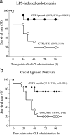
Therapeutic effect of sFlt-1 peptide on sepsis mortality. Survival curves for mice injected i.v. with sFlt-1 peptide or the same volume of PBS (control) 1 h after LPS injection (a) or CLP (b).

References

    1. Aird, W.C. 2003. The role of the endothelium in severe sepsis and multiple organ dysfunction syndrome. Blood. 101:3765–3777. - PubMed
    1. Angus, D.C., W.T. Linde-Zwirble, J. Lidicker, G. Clermont, J. Carcillo, and M.R. Pinsky. 2001. Epidemiology of severe sepsis in the United States: analysis of incidence, outcome, and associated costs of care. Crit. Care Med. 29:1303–1310. - PubMed
    1. Panacek, E.A., J.C. Marshall, T.E. Albertson, D.H. Johnson, S. Johnson, R.D. MacArthur, M. Miller, W.T. Barchuk, S. Fischkoff, M. Kaul, et al. 2004. Efficacy and safety of the monoclonal anti-tumor necrosis factor antibody F(ab')2 fragment afelimomab in patients with severe sepsis and elevated interleukin-6 levels. Crit. Care Med. 32:2173–2182. - PubMed
    1. Bernard, G.R., J.L. Vincent, P.F. Laterre, S.P. LaRosa, J.F. Dhainaut, A. Lopez-Rodriguez, J.S. Steingrub, G.E. Garber, J.D. Helterbrand, E.W. Ely, and C.J. Fisher Jr. 2001. Efficacy and safety of recombinant human activated protein C for severe sepsis. N. Engl. J. Med. 344:699–709. - PubMed
    1. Senger, D.R., S.J. Galli, A.M. Dvorak, C.A. Perruzzi, V.S. Harvey, and H.F. Dvorak. 1983. Tumor cells secrete a vascular permeability factor that promotes accumulation of ascites fluid. Science. 219:983–985. - PubMed

Publication types