

Moclobemide upregulated Bcl-2 expression and induced neural stem cell differentiation into serotoninergic neuron via extracellular-regulated kinase pathway
- PMID: 16702990
- PMCID: PMC1751873
- DOI: 10.1038/sj.bjp.0706766
Moclobemide upregulated Bcl-2 expression and induced neural stem cell differentiation into serotoninergic neuron via extracellular-regulated kinase pathway
Abstract
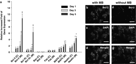
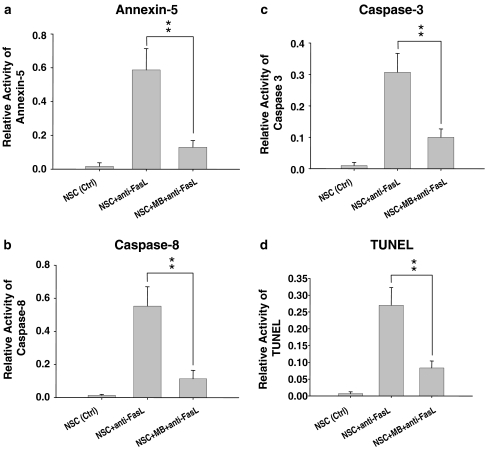
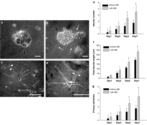
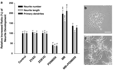
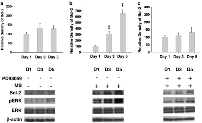
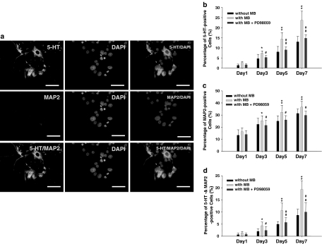
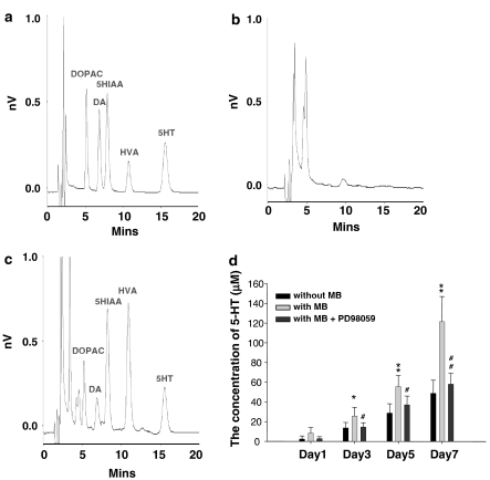
1. Moclobemide (MB) is an antidepressant drug that selectively and reversibly inhibits monoamine oxidase-A. Recent studies have revealed that antidepressant drugs possess the characters of potent growth-promoting factors for the development of neurogenesis and improve the survival rate of serotonin (5-hydroxytrytamine; 5-HT) neurons. However, whether MB comprises neuroprotection effects or modulates the proliferation of neural stem cells (NSCs) needs to be elucidated. 2. In this study, firstly, we used the MTT (3-(4,5-dimethylthiazol-2-yl)-2,5-diphenyltetrazolium bromide) assay to demonstrate that 50 microM MB can increase the cell viability of NSCs. The result of real-time reverse transcription-polymerase chain reaction (RT-PCR) showed that the induction of MB can upregulate the gene expressions of Bcl-2 and Bcl-xL. By using caspases 8 and 3, ELISA and terminal dUTP nick-end labeling (TUNEL) assay, our data further confirmed that 50 microM MB-treated NSCs can prevent FasL-induced apoptosis. 3. The morphological findings also supported the evidence that MB can facilitate the dendritic development and increase the neurite expansion of NSCs. Moreover, we found that MB treatment increased the expression of Bcl-2 in NSCs through activating the extracellular-regulated kinase (ERK) phosphorylation. 4. By using the triple-staining immunofluorescent study, the percentages of serotonin- and MAP-2-positive cells in the day 7 culture of MB-treated NSCs were significantly increased (P<0.01). Furthermore, our data supported that MB treatment increased functional production of serotonin in NSCs via the modulation of ERK1/2. In sum, the study results support that MB can upregulate Bcl-2 expression and induce the differentiation of NSCs into serotoninergic neuron via ERK pathway.
Figures

Comment in
-
Differentiation of hippocampal stem cells into functional neurons: evolving our understanding of monoamine oxidase-A inhibition.Br J Pharmacol. 2006 Jul;148(5):563-4. doi: 10.1038/sj.bjp.0706768. Epub 2006 May 15. Br J Pharmacol. 2006. PMID: 16702988 Free PMC article.
Similar articles
-
Antidepressant administration modulates neural stem cell survival and serotoninergic differentiation through bcl-2.Curr Neurovasc Res. 2007 Feb;4(1):19-29. doi: 10.2174/156720207779940707. Curr Neurovasc Res. 2007. PMID: 17311541
-
Neuroprotection by Imipramine against lipopolysaccharide-induced apoptosis in hippocampus-derived neural stem cells mediated by activation of BDNF and the MAPK pathway.Eur Neuropsychopharmacol. 2008 Feb;18(2):128-40. doi: 10.1016/j.euroneuro.2007.05.002. Epub 2007 Jun 12. Eur Neuropsychopharmacol. 2008. PMID: 17566715
-
Desipramine activated Bcl-2 expression and inhibited lipopolysaccharide-induced apoptosis in hippocampus-derived adult neural stem cells.J Pharmacol Sci. 2007 May;104(1):61-72. doi: 10.1254/jphs.fp0061255. J Pharmacol Sci. 2007. PMID: 17510525
-
Protective actions of ovarian hormones in the serotonin system of macaques.Front Neuroendocrinol. 2009 Jul;30(2):212-38. doi: 10.1016/j.yfrne.2009.04.003. Epub 2009 Apr 24. Front Neuroendocrinol. 2009. PMID: 19394356 Free PMC article. Review.
-
Driving apoptosis-relevant proteins toward neural differentiation.Mol Neurobiol. 2012 Oct;46(2):316-31. doi: 10.1007/s12035-012-8289-2. Epub 2012 Jul 1. Mol Neurobiol. 2012. PMID: 22752662 Review.
Cited by
-
Type A monoamine oxidase and serotonin are coordinately involved in depressive disorders: from neurotransmitter imbalance to impaired neurogenesis.J Neural Transm (Vienna). 2018 Jan;125(1):53-66. doi: 10.1007/s00702-017-1709-8. Epub 2017 Mar 14. J Neural Transm (Vienna). 2018. PMID: 28293733 Review.
-
Paroxetine up-regulates neurogenesis in hippocampus-derived neural stem cell from fetal rats.Mol Cell Biochem. 2013 Mar;375(1-2):105-13. doi: 10.1007/s11010-012-1533-2. Epub 2013 Jan 5. Mol Cell Biochem. 2013. PMID: 23291919
-
Evaluation of radiotherapy effect in resveratrol-treated medulloblastoma cancer stem-like cells.Childs Nerv Syst. 2009 May;25(5):543-50. doi: 10.1007/s00381-009-0826-6. Epub 2009 Feb 19. Childs Nerv Syst. 2009. PMID: 19225784
-
Neuroprotective effect of L-deprenyl on the expression level of the Mst1 gene and inhibition of apoptosis in rat-model spinal cord injury.Iran J Basic Med Sci. 2022 Jan;25(1):53-59. doi: 10.22038/IJBMS.2022.58031.12894. Iran J Basic Med Sci. 2022. PMID: 35656451 Free PMC article.
-
Neuroprotective effects of selegiline on rat neural stem cells treated with hydrogen peroxide.Biomed Rep. 2018 Jan;8(1):41-46. doi: 10.3892/br.2017.1023. Epub 2017 Nov 22. Biomed Rep. 2018. PMID: 29399337 Free PMC article.
References
-
- BACHMANN R.F., SCHLOESSER R.J., GOULD T.D., MANJI H.K.Mood stabilizers target cellular plasticity and resilience cascades: implications for the development of novel therapeutics Mol. Neurobiol. 200532173–202.Review - PubMed
-
- BONNET U., LENIGER T., WIEMANN M. Moclobemide reduces intracellular pH and neuronal activity of CA3 neurones in guinea-pig hippocampal slices – implication for its neuroprotective properties. Neuropharmacology. 2000;39:2067–2074. - PubMed
-
- CHEN D.F., SCHNEIDER G.E., MARTINOU J.C., TONEGAWA S. Bcl-2 promotes regeneration of severed axons in mammalian CNS. Nature. 1997;385:434–439. - PubMed
-
- CHENG F.C., KUO J.S., HUANG H.M., YANG D.Y., WU T.F., TSAI T.H. Determination of catecholamines in pheochromocytoma cell (PC-12) culture medium by microdialysis-microbore liquid chromatography. J. Chromatogr. A. 2000;870:405–411. - PubMed
Publication types
MeSH terms
Substances
LinkOut - more resources
Full Text Sources
Medical
Research Materials
Miscellaneous
