

Overexpression of the VAV proto-oncogene product is associated with B-cell chronic lymphocytic leukaemia displaying loss on 13q
- PMID: 16704440
- PMCID: PMC1950221
- DOI: 10.1111/j.1365-2141.2006.06094.x
Overexpression of the VAV proto-oncogene product is associated with B-cell chronic lymphocytic leukaemia displaying loss on 13q
Abstract
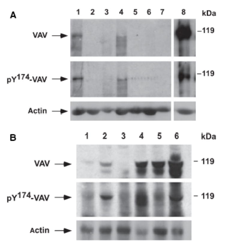
The expression of the VAV proto-oncogene in 57 patients with chronic myeloproliferative disease (CMD), B-cell acute lymphoblastic leukaemia (B-ALL) and B-cell non-Hodgkin Lymphoma (B-NHL), and 61 with B-cell chronic lymphocytic leukaemia (B-CLL) was analysed. VAV overexpression was observed in 19.5% of cases and 81% of VAV-positive tumours also displayed VAV phosphorylation. Overexpression was not observed in B-ALL or CMD, but 13% of B-NHL and 34.4% of B-CLL patients (P = 0.002) overexpressed VAV. The overexpression and phosphorylation of VAV was detected more frequently in 13q- chronic lymphocytic leukaemias (71.4%) versus other B-CLLs (23.4%, P = 0.001). Overexpression of VAV protein is a frequent event in patients with B-CLL displaying loss of 13q sequences.
Figures
References
-
- Etienne-Manneville S, Hall A. Rho GTPases in cell biology. Nature. 2002;420:629–635. - PubMed
-
- Fernández-Zapico ME, González-Paz NC, Weiss E, Savoy DN, Molina JR, Fonseca R, Smyrk TC, Chari ST, Urrutia R, Billadeau DD. Ectopic expression of VAV1 reveals an unexpected role in pancreatic cancer tumorigenesis. Cancer Cell. 2005;7:39–49. - PubMed
-
- Hernández JM, González MB, Granada I, Gutiérrez N, Chillón C, Ramos F, Ribera JM, González M, Feliu E, San Miguel JF. Detection of inv(16) and t(16;16) by fluorescence in situ hybridization in acute myeloid leukaemia M4Eo. Haematologica. 2000;85:481–485. - PubMed
