. 2006 May 23;103(21):8173-8.
doi: 10.1073/pnas.0510254103. Epub 2006 May 16.

The G12 family of heterotrimeric G proteins promotes breast cancer invasion and metastasis

Affiliations

The G12 family of heterotrimeric G proteins promotes breast cancer invasion and metastasis

Patrick Kelly et al. Proc Natl Acad Sci U S A. .

Abstract

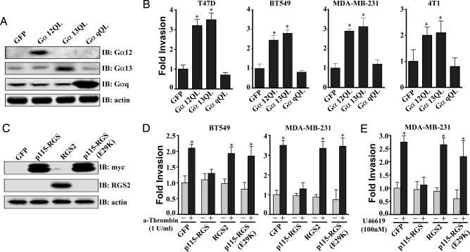
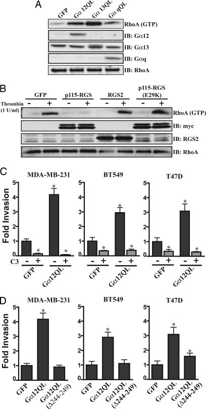
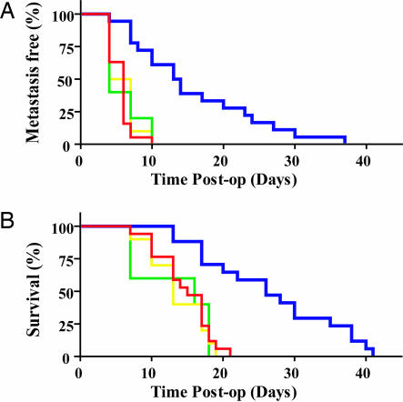
Although the prognosis for patients with early-stage breast cancer has improved, the therapeutic options for patients with locally advanced and metastatic disease are limited. To improve the treatment of these patients, the molecular mechanisms underlying breast cancer invasion and metastasis must be understood. In this study, we report that signaling through the G12 family of heterotrimeric G proteins (Galpha12 and Galpha13) promotes breast cancer cell invasion. Moreover, we demonstrate that inhibition of G12 signaling reduces the metastatic dissemination of breast cancer cells in vivo. Finally, we demonstrate that the expression of Galpha12 is significantly up-regulated in the earliest stages of breast cancer, implying that amplification of G12 signaling may be an early event in breast cancer progression. Taken together, these observations identify the G12 family proteins as important regulators of breast cancer invasion and suggest that these proteins may be targeted to limit invasion- and metastasis-induced patient morbidity and mortality.


Conflict of interest statement

Conflict of interest statement: No conflicts declared.

Figures

Fig. 1.
Fig. 1.
G12 signaling promotes breast cancer cell invasion. (A) Expression of the activated forms of Gα12 (Gα12QL) and Gα13 (Gα13QL) but not Gαq (GαqQL) induces breast cancer cell invasion in vitro. Cells were transduced with the indicated adenovirus, starved for 18 h, and then allowed to invade growth-factor reduced Matrigel (T47D, MDA-MB-231, and BT549) or collagen-coated (4T1) transwell filters for 30 h. (B) Immunoblot analysis showing expression levels of Gα12, Gα13, and Gαq in the T47D cell line after infection with the indicated adenovirus. Similar expression was observed in the other cells types. (C and D) Expression of p115-RGS blocks thrombin (C) and thromboxane A2-induced (U46619) (D) invasion of breast cancer cells, whereas expression of RGS2 or expression of an inactive form of the p115-RGS, p115-RGS(E29K), has no effect. Cells were transduced with the indicated adenovirus, starved for 18 h, then allowed to invade growth-factor-reduced Matrigel-coated transwell filters for 30 h in the presence of 1 unit/ml thrombin (A), 100 nM U46619 (B), or vehicle control. (E) Immunoblot analysis showing expression levels of p115-RGS, p115-RGS(E29K), and RGS2 in the MDA-MB-231 cell line after infection with the indicated adenovirus. Similar expression was observed in the BT549 cells. (A, C, and D) Experiments were performed in triplicate, and the results are presented as the fold increase over vehicle-treated GFP control. All results are presented as mean ± SE from a single experiment. All experiments were performed at least three times. ∗, P < 0.05 as determined by paired Student t test.
Fig. 2.
Fig. 2.
G12 signaling is able to promote breast cancer cell invasion through the activation of the Rho family of monomeric G proteins. (A) Expression of Gα12QL and Gα13QL induces RhoA activation in the MDA-MB-231 cell line. Cells were transduced with the indicated adenovirus and then starved for 18 h. Cells were lysed, and the lysates were subjected to pull-down assays using a GST fusion of the activated RhoA-binding domain of rhotekin. Levels of precipitated RhoA were determined by immunoblot analysis using anti-RhoA antibody. Levels of total RhoA, Gα12, Gα13, and Gαq also were determined. All lanes are representative of two or more separate experiments. (B) Thrombin-stimulated RhoA activation in MDA-MB-231 cells is inhibited by expression of p115-RGS but not by expression of RGS2 or p115-RGS(E29K). Cells were transduced with the indicated adenovirus, starved for 18 h, and then stimulated with thrombin (1 unit/ml) or a vehicle control for 5 min. Levels of activated Rho were determined as in A. Levels of total RhoA, myc-p115-RGS, and RGS2 also were determined. All lanes are representative of two or more separate experiments. (C) Gα12-mediated breast cancer cell invasion requires the activity of the Rho family of G proteins. Cells were transduced with the indicated adenovirus, starved for 18 h in the presence and absence of C3 toxin as indicated, and then allowed to invade growth-factor-reduced Matrigel-coated transwell filters for 30 h. Experiments were performed in duplicate, and the results are presented as the fold increase over that observed with GFP control. All experiments were performed at least three times. All results are presented as mean ± SE. ∗, P < 0.05 as determined by paired Student t test. (D) The effect of the expression of Gα12QL (Δ244–249), a mutant of Gα12QL that is functionally uncoupled from the Rho axis but is still able to interact with other putative G12 effectors, on breast cancer cell invasion. Experiments were performed as in C. Experiments were preformed in duplicate, and the results are presented as the fold increase over that observed with GFP control. All experiments were performed at least three times. All results are presented as mean ± SE. ∗, P < 0.05 as determined by paired Student t test.
Fig. 3.
Fig. 3.
Inhibition of G12 signaling reduces metastatic dissemination of the 4T1 cell line orthotopically implanted in the mouse mammary fat pad. The mammary fat pads of BALB/c mice were injected with 5 × 10⁵ 4T1-Luc cells (red; n = 17), 4T1-Luc cells expressing p115-RGS (blue; n = 16), 4T1-Luc cells expressing RGS2 (yellow; n = 11), or 4T1-Luc cells expressing p115-RGS(E29K) (green; n = 5). Tumors were grown for 21 days and then excised as described in Methods and Materials. (A) Metastasis-free survival after primary tumor resection. A metastatic event was defined as any detectable bioluminescent signal above background away from the primary site. Mice that received p115-RGS-expressing 4T1 cells had significantly longer metastasis-free survival times than did mice that received control 4T1 cells (P < 0.0001), RGS2-expressing cells (P < 0.001), or p115-RGS(E29K)-expressing cells (P < 0.0013). Statistical analyses were performed by using the log-rank test. (B) Overall mouse survival. Mice that received p115-RGS-expressing 4T1 cells survived significantly longer than did mice that received control 4T1 cells (P < 0.0001), RGS2-expressing cells (P < 0.001), or p115-RGS(E29K)-expressing cells (P < 0.002). Statistical analyses were performed by using the log-rank test.
Fig. 4.
Fig. 4.
Gα12 protein levels are up-regulated in carcinoma in situ and in invasive adenocarcinoma of the breast. Sections of formalin-fixed paraffin-embedded breast tissue were stained for Gα12 with anti-G12 anti-sera (Santa Cruz Biotechnology) as described in Methods and Materials. Original images were taken with a ×40 objective. (A and B) Benign breast tissue. (C) Ductal carcinoma in situ of the breast. (D) Lobular carcinoma in situ of the breast. (E) Section containing benign breast epithelium, carcinoma in situ, and invasive ductal carcinoma of the breast. (F) Section containing both benign breast epithelium and invasive lobular carcinoma of the breast. (E and F) Arrowheads indicate cancer; arrows indicate benign epithelium.

References

    1. Pierce K. L., Premont R. T., Lefkowitz R. J. Nat. Rev. Mol. Cell. Biol. 2002;3:639–650. - PubMed
    1. Rohrer D. K., Kobilka B. K. Physiol. Rev. 1998;78:35–52. - PubMed
    1. Offermanns S. Oncogene. 2001;20:1635–1642. - PubMed
    1. Strathmann M. P., Simon M. I. Proc. Natl. Acad. Sci. USA. 1991;88:5582–5586. - PMC - PubMed
    1. Chan A. M., Fleming T. P., McGovern E. S., Chedid M., Miki T., Aaronson S. A. Mol. Cell. Biol. 1993;13:762–768. - PMC - PubMed

Publication types

Substances