. 2006 Jun 1;20(11):1470-84.
doi: 10.1101/gad.1416106. Epub 2006 May 16.

The Evf-2 noncoding RNA is transcribed from the Dlx-5/6 ultraconserved region and functions as a Dlx-2 transcriptional coactivator

Affiliations

The Evf-2 noncoding RNA is transcribed from the Dlx-5/6 ultraconserved region and functions as a Dlx-2 transcriptional coactivator

Jianchi Feng et al. Genes Dev. .

Abstract

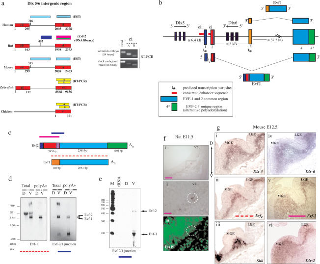
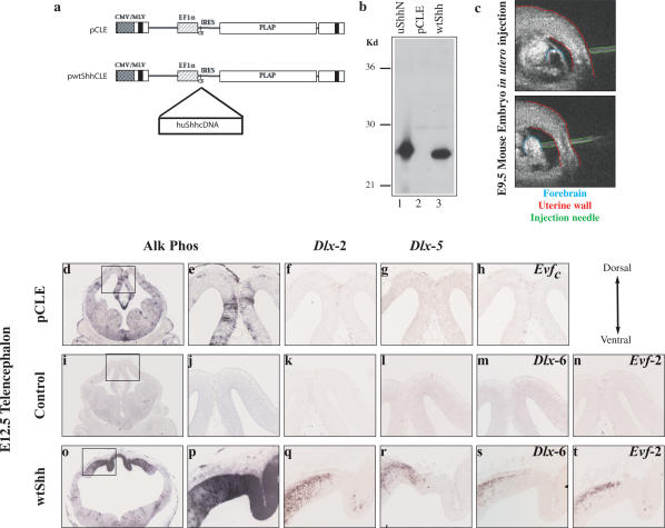
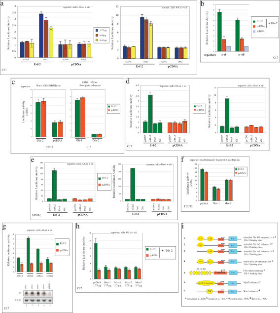
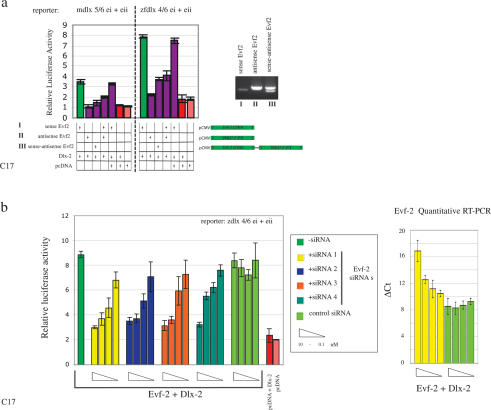
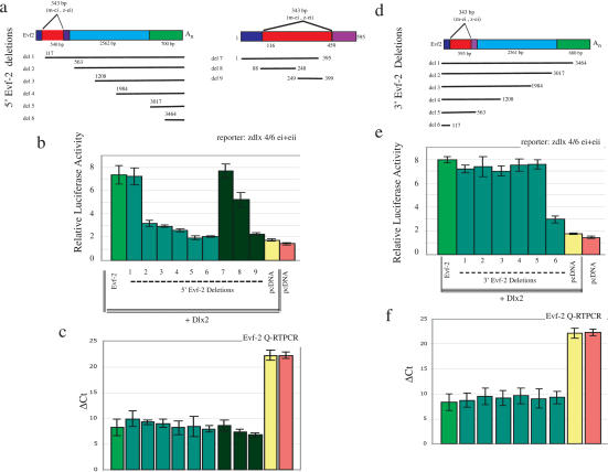
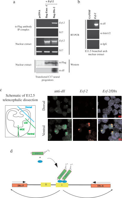
The identification of ultraconserved noncoding sequences in vertebrates has been associated with developmental regulators and DNA-binding proteins. One of the first of these was identified in the intergenic region between the Dlx-5 and Dlx-6 genes, members of the Dlx/dll homeodomain-containing protein family. In previous experiments, we showed that Sonic hedgehog treatment of forebrain neural explants results in the activation of Dlx-2 and the novel noncoding RNA (ncRNA), Evf-1. In this report, we show that the Dlx-5/6 ultraconserved region is transcribed to generate an alternatively spliced form of Evf-1, the ncRNA Evf-2. Evf-2 specifically cooperates with Dlx-2 to increase the transcriptional activity of the Dlx-5/6 enhancer in a target and homeodomain-specific manner. A stable complex containing the Evf-2 ncRNA and the Dlx-2 protein forms in vivo, suggesting that the Evf-2 ncRNA activates transcriptional activity by directly influencing Dlx-2 activity. These experiments identify a novel mechanism whereby transcription is controlled by the cooperative actions of an ncRNA and a homeodomain protein. The possibility that a subset of vertebrate ultraconserved regions may function at both the DNA and RNA level to control key developmental regulators may explain why ultraconserved sequences exhibit 90% or more conservation even after 450 million years of vertebrate evolution.


Figures

Figure 1.
Figure 1.
Identification of Evf-2 transcripts. (a) The ultraconserved Dlx-5/6 intergenic enhancer ei is transcribed in vertebrates. Human and mouse ESTs were found in the database (sky blue). ei-containing ESTs were not found in the zebrafish and chick databases. The right panel shows that two different primer sets (A and B) detect ei-containing transcripts in cDNAs made from zebrafish embryos or chick embryonic brains. (b) Genomic organization of the mouse Evf gene. Evf-1 and Evf-2 result from alternate transcription initiation, alternative splicing of exon 3, and alternative polyadenylation. Exon 2, included in Evf-2 but absent from Evf-1, is partly encoded by the conserved intergenic enhancer ei (red). (c) Schematic representation of Evf-1 and Evf-2. Common regions are shown in blue. Evf-2 nucleotides 116–459 are encoded by the conserved intergenic enhancer, ei (red). Antisense RNA probes (shown as pink, blue, and red dashed lines) spanning different regions of Evf-1 and Evf-2 were used for Northern, RNase protection, and in situ hybridizations, as indicated. (d) Northern analysis of total and polyadenylated RNAs isolated from rat E13.5 embryonic telencephalon, using probes generated against Evf-1 and Evf-2 (as indicated in c). (D) Dorsal; (V) ventral. The full-length Evf-1 probe detects the 28S rRNA in total RNAs from both dorsal and ventral RNAs, showing that the absence of Evfs in D RNAs is not due to degradation. (e) RNase protection. A 450-nt probe spanning the Evf 2/common region junction (blue) detects RNase-resistant bands in ventral (V) but not in dorsal (D) rat E13.5 embryonic telencephalic RNAs or tRNA. (f, panels i,ii) Section in situ hybridization of tissues from rat ventral forebrain at E11.5 using the 5′specific Evf-2 probe (pink). (Panel iii) Dense Evf-2 staining colocalizes with the nuclear stain DAPI. (g) Section in situ hybridization of mouse ventral forebrain at E12.5 using the following antisense probes: Dlx-5 (panel i), Evf (1 + common region) (panel ii), Shh (panel iii), Dlx-6 (panel iv), Evf-2 (panel v), and Dlx-2 (panel vi). (VZ) Ventricular zone; (LGE) lateral ganglionic eminence; (MGE) medial ganglionic eminence; (D) dorsal; (V) ventral. The full-length rat Evf-1 cDNA sequence is available on GenBank (accession no. AY518691).
Figure 2.
Figure 2.
Sonic hedgehog induces the expression of Evf, Dlx-2, and Dlx-5 in vivo. (a) Diagram of retroviral backbone used to express the human sonic hedgehog protein (pwtShhCLE) or control (pCLE). Alkaline phosphatase is bicistronic with the Shh cDNA, allowing detection of virally infected cells. (b) Lysates of C17 cells infected with wtShh virus (lane 3), pCLE control virus (lane 2), or 7 ng of purified recombinant-unmodified Shh (lane 1, uShhN) were Western-blotted and probed with anti-Shh antibody (Santa Cruz Biotechnology). (c) In utero UBM-guided entry of viruses into E9.5 mouse forebrain. Sections of E12.5 mouse brains 3 d after infection with control pCLE virus (d–h), littermate control (i–n), or wtShh virus (o–t). (d,e) pCLE virus-infected clusters. (o,p) wtshh-infected viral clusters visualized by alkaline phosphatase staining in the dorsal midline of the telencephalon. (i,j) Uninfected littermate control does not contain alkaline phosphatase-expressing clusters of cells. In situ hybridization of adjacent sections probed for ectopic expression of ventral genes Dlx-2 (f,k,q), Dlx-5 (g,l,r), Evfc (h), Dlx-6 (m,s), and Evf-2 (n,t). Orientation of the section is indicated in the top right box.
Figure 3.
Figure 3.
Evf-2 cooperates with Dlx family members to activate Dlx-5/6 enhancer activity in a target-specific, homeodomain-specific, and cell-type-specific manner. The C17 and MN9D neural cell lines or the C2C12 muscle cell line, as indicated, were transfected with pcDNA-containing constructs along with the different reporters, as indicated (see i). All experiments were performed a minimum of three times in triplicate. The different reporters listed in i 1–7 were used as targets to determine the specificity required for cooperative activation by Evf-2 and Dlx-2. Transfection efficiency was normalized by including a Renilla luciferase internal control plasmid. (a) Evf-2 induces dosage-dependent cooperative activation of the mouse Dlx-5/6 and zebrafish Dlx-4/6 enhancer constructs. Evf-2 (1.75, 0.88, or 0.44 μg) was cotransfected with pcDNA–Dlx-2, along with different reporter constructs, and Firefly and Renilla luciferase activities were determined, normalized, and plotted on the Y-axis. (b) Both ei and eii are targets of Evf-2 transcription-enhancing activity. (c) Evf-2 does not cooperate with Dlx-2 to increase the activation of the Wnt enhancer and does not cooperate with Gli-1 to increase the activation of the floor plate enhancer. (d) Evf-2 does not cooperate with Pax3 or Gli-1 to activate Dlx-5/6 enhancer activity in C17 neural cells. (e) Evf-2 cooperates with Dlx-2 but not with Pax-3 or Gli-1 to activate Dlx-5/6 enhancer activity in MN9D neural cells. (f) Evf-2 does not cooperate with Msx-1 and Msx-2 to suppress the myoD enhancer in the muscle cell line C2C12. (g) Dlx family members 1, 2, 4, and 6 exhibit cooperative activity with Evf-2 to different levels, Dlx-2 > Dlx-4 > Dlx-6 > Dlx-1. Western analysis of transfected cell extracts probed with pan-anti-dll antibody is shown below. (h) Evf-2 does not prevent inhibition by Msx-1 and Msx-2. (i) Summary of reporters used in ah.
Figure 4.
Figure 4.
The active form of Evf-2 is single-stranded RNA. (a) The sense (I), but not antisense (II) or double-stranded (III) form of Evf-2 cooperates with Dlx-2 to increase the transcriptional activity of the Dlx-5/6 enhancer. Antisense decreases the activity of the sense form. RT–PCR of C17 cells transfected with constructs I, II, and III show that all three forms are made. (b) siRNAs (1–4) directed against Evf-2 inhibit Evf-2/Dlx-2 transcriptional activation in a dose-dependent manner. siRNA-1 (yellow bars) degrades Evf-2 RNA levels as assessed by quantitative RT–PCR, whereas a control siRNA generated against luciferase (green bars) does not. (ΔCt) Number of Evf-2 cycles minus the number of β-actin cycles, repeated three times and averaged.
Figure 5.
Figure 5.
The Evf-2 5′ region is both necessary and sufficient for Dlx-2-dependent cooperative activation of the Dlx-5/6 enhancer. Comparison of Dlx-5/6 enhancer activation in C17 neural cell lines by different Evf-2 deletion mutants. (a) Diagram of 5′ Evf-2 deletions. (b) Activity of different 5′ Evf-2 deletions. (c) Quantitative RT–PCR of Evf-2 deletions. (d) Diagram of 3′ Evf-2 deletions. (e) Activity of different 3′ Evf-2 deletions. (f) Quantitative RT–PCR of 3′ Evf-2 deletions. Quantitative RT–PCR analysis shows that 5′ and 3′ deletions are made in relatively similar amounts. Therefore, loss of function is not the result of Evf-2 RNA degradation. (ΔCt) Number of Evf-2 cycles minus the number of β-actin cycles, repeated three times and averaged.
Figure 6.
Figure 6.
Evf-2 and Dlx-2 form a complex in vivo. (a) Nuclear extracts made from C17 neural cells transfected with Flag-tagged Emx-1, Flag-tagged Dlx-2, or pcDNA control were analyzed for the presence of Evf-2–Dlx-2 complexes by immunoprecipitation with anti-Flag antibody, followed by RT–PCR against Evf-2-specific primers and S17 control primers. Western analysis shows that both Flag-Emx-1 and Flag-Dlx-2 are present in nuclear extracts transfected with constructs expressing these proteins. (b) Nuclear extracts made from rat E11.5 BAs were analyzed for the presence of Evf-2/Dlx-2 complexes by immunoprecipitation with anti-dll, anti-Islet 1/2, or anti-IgG antibody, followed by RT–PCR against Evf-2-specific primers or GAPDH. (c) Single-cell suspensions made from mouse E12.5 dorsal and ventral telencephalon were dissected as shown in the schematic, centrifuged onto slides, and processed for fluorescent in situ/immunolocalization using Evf-2 antisense RNA probe and anti-dll antibody. DAPI staining (blue) reveals nuclei. Evf-2 RNA (Alexa fluor 568) is in red, Dlx (Alexa fluor 488) is in green, and regions of overlap are in yellow. (d) A model proposing that a complex of Evf-2 and Dlx-2 affects ei activity, ultimately affecting transcription of the Dlx-5 and Dlx-6 genes.

References

    1. Acampora D., Merlo G.R., Paleari L., Zerega B., Postiglione M.P., Mantero S., Bober E., Barbieri O., Simeone A., Levi G. Craniofacial, vestibular and bone defects in mice lacking the Distal-less related gene. Dlx5. Development. 1999;126:3795–3809. - PubMed
    1. Akhtar A. Dosage compensation: An intertwined world of RNA and chromatin remodeling. Curr. Opin. Genet. Dev. 2003;13:161–169. - PubMed
    1. Amrein H., Axel R. Genes expressed in neurons of adult male Drosophila. Cell. 1997;88:459–469. - PubMed
    1. Anderson S., Eisenstat D., Shi L., Rubenstein J. Interneuron migration from basal forebrain to neocortex: Dependence on Dlx genes. Science. 1997a;278:474–476. - PubMed
    1. Anderson S.A., Qiu M., Bulfone A., Eisenstat D.D., Meneses J., Pedersen R., Rubenstein J.L.R. Mutations of the homeobox genes Dlx-1 and Dlx-2 disrupt the striatal subventricular zone and differentiation of late born striatal neurons. Neuron. 1997b;19:27–37. - PubMed

Publication types