. 2006 Jun 1;20(11):1496-510.
doi: 10.1101/gad.1417406. Epub 2006 May 16.

Lung adenocarcinomas induced in mice by mutant EGF receptors found in human lung cancers respond to a tyrosine kinase inhibitor or to down-regulation of the receptors

Affiliations

Lung adenocarcinomas induced in mice by mutant EGF receptors found in human lung cancers respond to a tyrosine kinase inhibitor or to down-regulation of the receptors

Katerina Politi et al. Genes Dev. .

Abstract

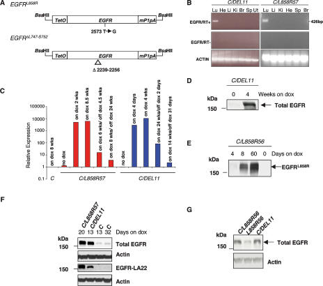
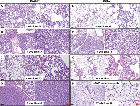
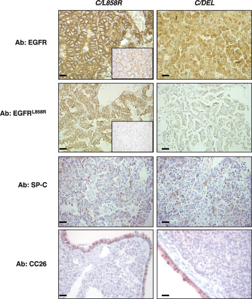
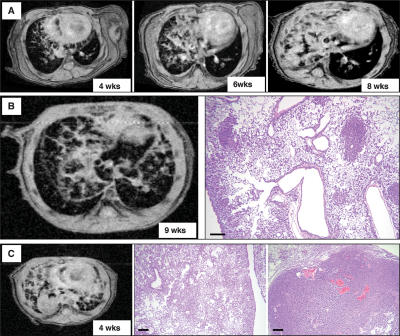
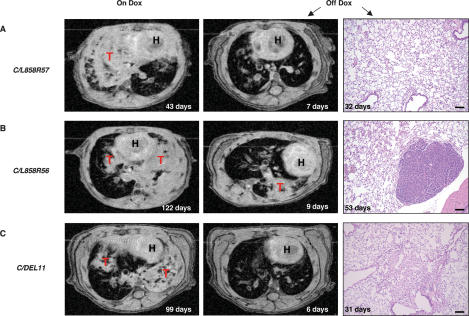
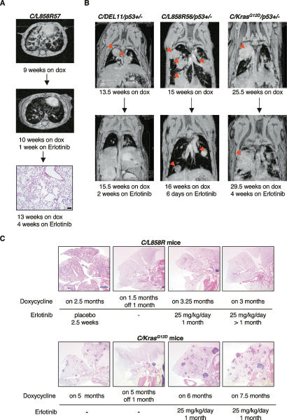
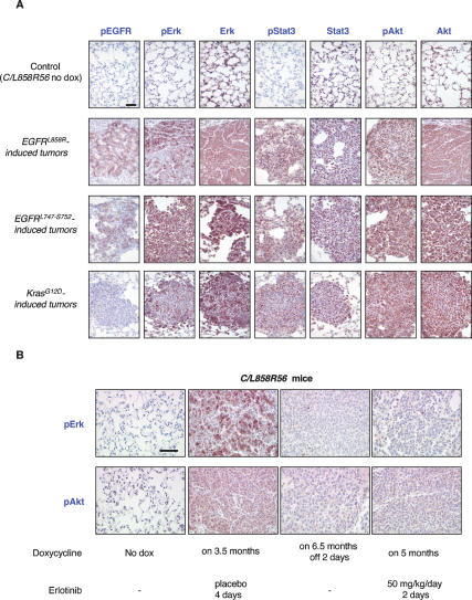
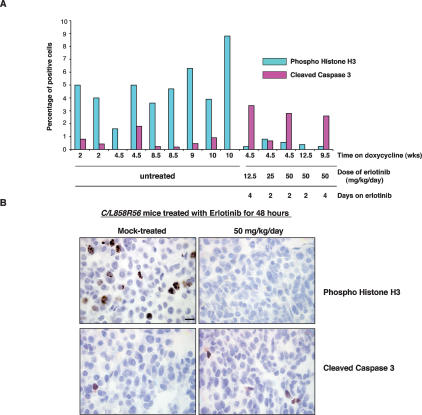
Somatic mutations in exons encoding the tyrosine kinase domain of the epidermal growth factor receptor (EGFR) gene are found in human lung adenocarcinomas and are associated with sensitivity to the tyrosine kinase inhibitors gefitinib and erlotinib. Nearly 90% of the EGFR mutations are either short, in-frame deletions in exon 19 or point mutations that result in substitution of arginine for leucine at amino acid 858 (L858R). To study further the role of these mutations in the initiation and maintenance of lung cancer, we have developed transgenic mice that express an exon 19 deletion mutant (EGFR(DeltaL747-S752)) or the L858R mutant (EGFR(L858R)) in type II pneumocytes under the control of doxycycline. Expression of either EGFR mutant leads to the development of lung adenocarcinomas. Two weeks after induction with doxycycline, mice that express the EGFR(L858R) allele show diffuse lung cancer highly reminiscent of human bronchioloalveolar carcinoma and later develop interspersed multifocal adenocarcinomas. In contrast, mice expressing EGFR(DeltaL747-S752) develop multifocal tumors embedded in normal lung parenchyma with a longer latency. With mice carrying either EGFR allele, withdrawal of doxycycline (to reduce expression of the transgene) or treatment with erlotinib (to inhibit kinase activity) causes rapid tumor regression, as assessed by magnetic resonance imaging and histopathology, demonstrating that mutant EGFR is required for tumor maintenance. These models may be useful for developing improved therapies for patients with lung cancers bearing EGFR mutations.


Figures

Figure 1.
Figure 1.
EGFR transgenes and their expression patterns. (A) Constructs used to generate tetracycline-inducible EGFR transgenic mice. Mutant human EGFR cDNAs (EGFRL858R and EGFRΔL747–S752) were cloned between a tetracycline operator sequence and the mouse protamine-1 polyadenylation sequence. (TetO) Tetracycline operator; (mP1pA) mouse protamine-1 gene polyadenylation sequence. (B) Expression of EGFRΔL747–S752 (DEL11, left panels) and EGFRL858R (L858R57, right panels) is restricted to lung tissue in bitransgenic mice maintained on doxycycline for 8 wk and 3 wk, respectively. PCR reactions were carried out in the presence (top) and absence (middle) of RT and products were visualized after electrophoresis in a 2% agarose gel. Amplification of actin mRNA by RT–PCR confirmed the presence of RNA in all samples. (Lu) Lung; (He) heart; (Li) liver; (Ki) kidney; (Br) brain; (Sp) spleen; (Ut) uterus. (C) Quantitative RT–PCR analysis of transgene expression in C/L858R57 and C/DEL11 bitransgenic mice and C transgenic control mice before, during, and after doxycycline treatment. (DG) Mutant EGFR protein is expressed at high levels in lungs from induced mice. (D) Lung extracts from C/DEL11 mice maintained on doxycycline for the times indicated were immunoblotted with an antibody that recognizes human and mouse EGFR (Total EGFR). (E) Lung protein extracts from C/L858R bitransgenic mice maintained on doxycycline for the times indicated were immunoblotted with an EGFRL858R-specific antibody (see Materials and Methods for details). (F) Western analysis of lung protein extracts from mice maintained on doxycycline for 13–32 d using the total EGFR antibody and an antibody that recognizes human EGFR with a higher affinity than mouse EGFR (EGFR-LA22, third panel). (G) Western analysis of lung protein extracts from bitransgenic mice maintained on doxycycline for 4 d using the total EGFR antibody. Anti-actin blots are shown as loading controls.
Figure 2.
Figure 2.
C/L858R and C/DEL mice fed doxycycline develop lung tumors. Hematoxylin and eosin (H&E)-stained sections of lung samples derived from C/L858R (AD) and C/DEL (EG) bitransgenic mice after treatment with doxycycline for the indicated times. Labels indicate the transgenic EGFR line and time on doxycycline. C and D contain images derived from the same lung, illustrating different pathology in different regions. (H) Lung tissue from a doxycycline-treated L858R57 monotransgenic mouse after prolonged treatment with doxycyline. Black bars, 100 μm. Red bars, 20 μm.
Figure 3.
Figure 3.
Lung tumors induced in C/L858R and C/DEL mice contain abundant EGFR protein and are composed mainly of type II pneumocytes. Sections were stained with antibodies to total EGFR, EGFRL858R, SP-C, and CC26 as indicated. The insets in samples from EGFRL858R-induced tumors show the tissue stained without the primary antibody. Bars, 20 μm.
Figure 4.
Figure 4.
Lung tumor growth can be monitored by MRI. (A) Serial MRI of a C/L858R57 mouse after 4, 6, and 8 wk on doxycycline shows a pattern consistent with a diffuse reticulonodular infiltrate in the right lung that progressively increased in size and became more consolidated. (B, left) MR image of the lungs from a C/L858R57 mouse fed doxycycline for 9 wk. (Right) The reticulonodular pattern observed by MRI reflects diffuse BAC and tumor nodules throughout the affected lung field, as shown in the H&E-stained section. (C, left) MR image of the lungs from a C/L858R56 bitransgenic mouse fed doxycycline for 4 wk showing reticulonodular consolidation and a large solid nodule. These features on MRI correspond to diffuse BAC (middle) and an invasive adenocarcinoma (right) observed histologically. Bars, 200 μm.
Figure 5.
Figure 5.
Lung opacities disappear in MR images, and the tumors regress histologically in the lungs of bitransgenic mice after withdrawal of doxycycline. (A–C) Serial MR images from the lungs of C/L858R and C/DEL mice maintained on doxycycline for the times indicated (left panels) and after deinduction (middle panels). (Right panels) H&E-stained sections from the mice after deinduction for the times indicated on the panels. (H) Heart; (T) tumor. Bars, 100 μm (see Supplementary Table 3 for additional information on these mice, and the text for a discussion of the peristent nodule shown in B, right panel).
Figure 6.
Figure 6.
Lung tumors in C/L858R or C/DEL, but not C/KrasG12D bitransgenic mice on doxycycline disappear during treatment with erlotinib. (A) Axial MR images of lungs from a C/L858R57 mouse after 9 wk on doxycycline (top) and after 1 wk of erlotinib treatment (25 mg/kg/d) plus continued doxycycline (middle). The mouse was sacrificed after 4 wk of erlotinib treatment, and an H&E-stained section of the lung is shown in the bottom panel. Black bar, 100 μm. (B) Coronal MR images of the lungs of C/DEL11/p53+/− (left), C/L858R56/p53+/− (center), and C/KrasG12D/p53+/− (right) mice before (top) and after (bottom) receiving erlotinib for the indicated times. Red arrows indicate tumors. (C) H&E-stained lung sections from C/L858R (top) and C/KrasG12D (bottom) mice, placebo-treated, erlotinib-treated, or deinduced as indicated. These images provide a qualitative, but not quantitative, assessment of the effect of erlotinib on mice that express oncogenic Kras. A quantitative assessment would require knowledge of the initial tumor burden in individual mice prior to erlotinib therapy. Blue bar, 2 mm.
Figure 7.
Figure 7.
Multiple signaling pathways are activated in mutant EGFR and KrasG12D-induced lung tumors and deactivated during tumor regression. (A) Immunohistochemical staining was used to detect pEGFR, pErk, Erk, pStat3, Stat3, pAkt, and Akt in L858R-, DEL-, and KrasG12D-induced lung tumors and in uninduced lungs (normal lung tissue derived from a bitransgenic C/L858R56 mouse maintained on normal chow was stained as a control) as indicated. (B) Immunohistochemical staining was used to detect pErk and pAkt in lung tumors from C/L858R56 mice after 2 d of erlotinib treatment or doxycycline withdrawal and in uninduced and placebo-treated lungs as indicated. Bar, 50 μm.
Figure 8.
Figure 8.
Decreased mitoses and increased apoptosis upon effective erlotinib treatment. (A) Quantification of phosphohistone H3 and cleaved caspase 3 in untreated and erlotinib-treated mice. (B) Phosphohistone H3 (top) and cleaved caspase 3 (bottom) staining of lung sections from untreated and erlotinib-treated C/L858R56 mice is shown. Bar, 10 μm.

References

    1. Amann J., Kalyankrishna S., Massion P.P., Ohm J.E., Girard L., Shigematsu H., Peyton M., Juroske D., Huang Y., Stuart Salmon J., et al. Aberrant epidermal growth factor receptor signaling and enhanced sensitivity to EGFR inhibitors in lung cancer. Cancer Res. 2005;65:226–235. - PubMed
    1. Andrechek E.R., Hardy W.R., Siegel P.M., Rudnicki M.A., Cardiff R.D., Muller W.J. Amplification of the neu/erbB-2 oncogene in a mouse model of mammary tumorigenesis. Proc. Natl. Acad. Sci. 2000;97:3444–3449. - PMC - PubMed
    1. Barkley J.E., Green M.R. Bronchioloalveolar carcinoma. J. Clin. Oncol. 1996;14:2377–2386. - PubMed
    1. Boxer R.B., Jang J.W., Sintasath L., Chodosh L.A. Lack of sustained regression of c-MYC-induced mammary adenocarcinomas following brief or prolonged MYC inactivation. Cancer Cell. 2004;6:577–586. - PubMed
    1. Cappuzzo F., Hirsch F.R., Rossi E., Bartolini S., Ceresoli G.L., Bemis L., Haney J., Witta S., Danenberg K., Domenichini I., et al. Epidermal growth factor receptor gene and protein and gefitinib sensitivity in non-small-cell lung cancer. J. Natl. Cancer Inst. 2005;97:643–655. - PubMed

Publication types